Int J Mol Sci. 2021 Oct 13;22(20):11030. doi: 10.3390/ijms222011030 https://pmc.ncbi.nlm.nih.gov/articles/PMC8538579/
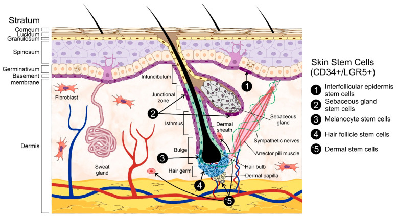
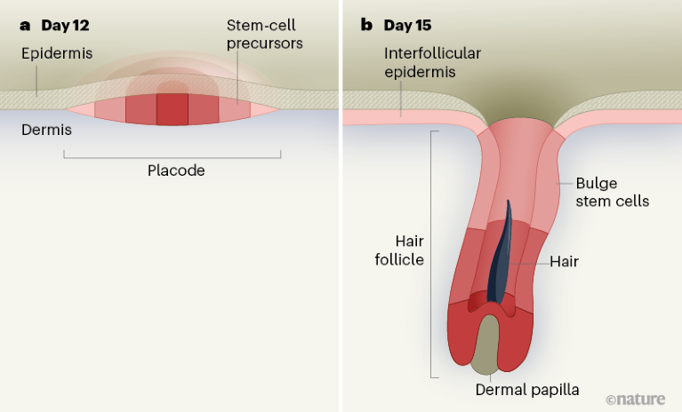
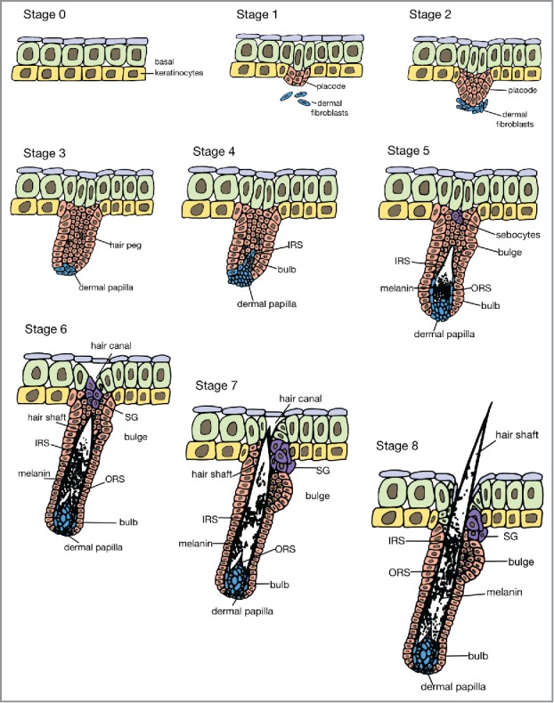
毛の形成
間葉が表皮に働きかけて、表皮の一部がプラコード(原基)に分化します。
09 June 2021 A 4D road map for the formation of hair follicles https://www.nature.com/articles/d41586-021-01482-1
Organogenesis. 2018 Feb 15;14(1):46–63. doi: 10.1080/15476278.2017.1421882 Embryonic skin development and repair https://pmc.ncbi.nlm.nih.gov/articles/PMC6150059/
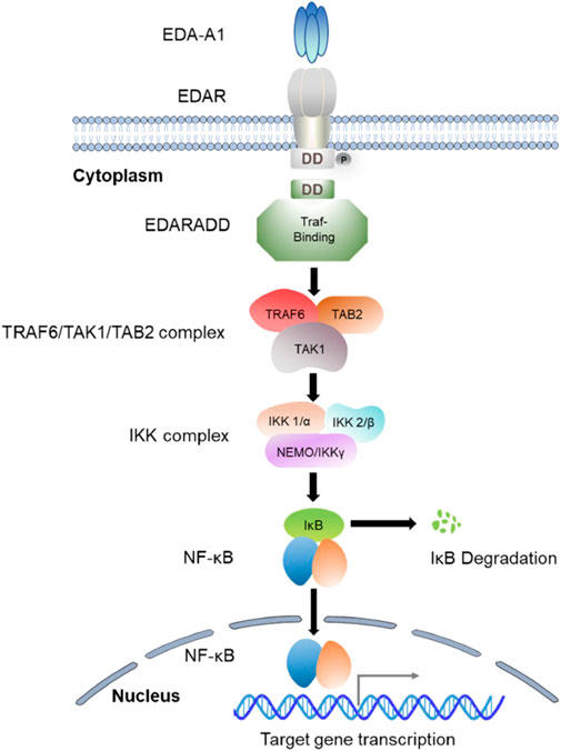
MINI REVIEW article Front. Genet., 03 April 2023 Volume 14 – 2023 | https://doi.org/10.3389/fgene.2023.1168538 The EDA/EDAR/NF-κB pathway in non-syndromic tooth agenesis: A genetic perspective https://www.frontiersin.org/journals/genetics/articles/10.3389/fgene.2023.1168538/full
毛の発生のシグナリング
Nature Communications volume 9, Article number: 2333 (2018) Published: 20 June 2018 FGF signalling controls the specification of hair placode-derived SOX9 positive progenitors to Merkel cells
STEM CELLS Tissue-Specific Stem Cells Free Access Concise Review: Wnt Signaling Pathways in Skin Development and Epidermal Stem Cells Anthony Veltri, Christopher Lang, Wen-Hui Lien First published: 19 October 2017 https://doi.org/10.1002/stem.2723 https://stemcellsjournals.onlinelibrary.wiley.com/doi/10.1002/stem.2723
毛の再生シグナリング
Front Med (Lausanne). 2022 Apr 14;9:863786. doi: 10.3389/fmed.2022.863786 Epithelial-Mesenchymal Interaction in Hair Regeneration and Skin Wound Healing https://pmc.ncbi.nlm.nih.gov/articles/PMC9048199/
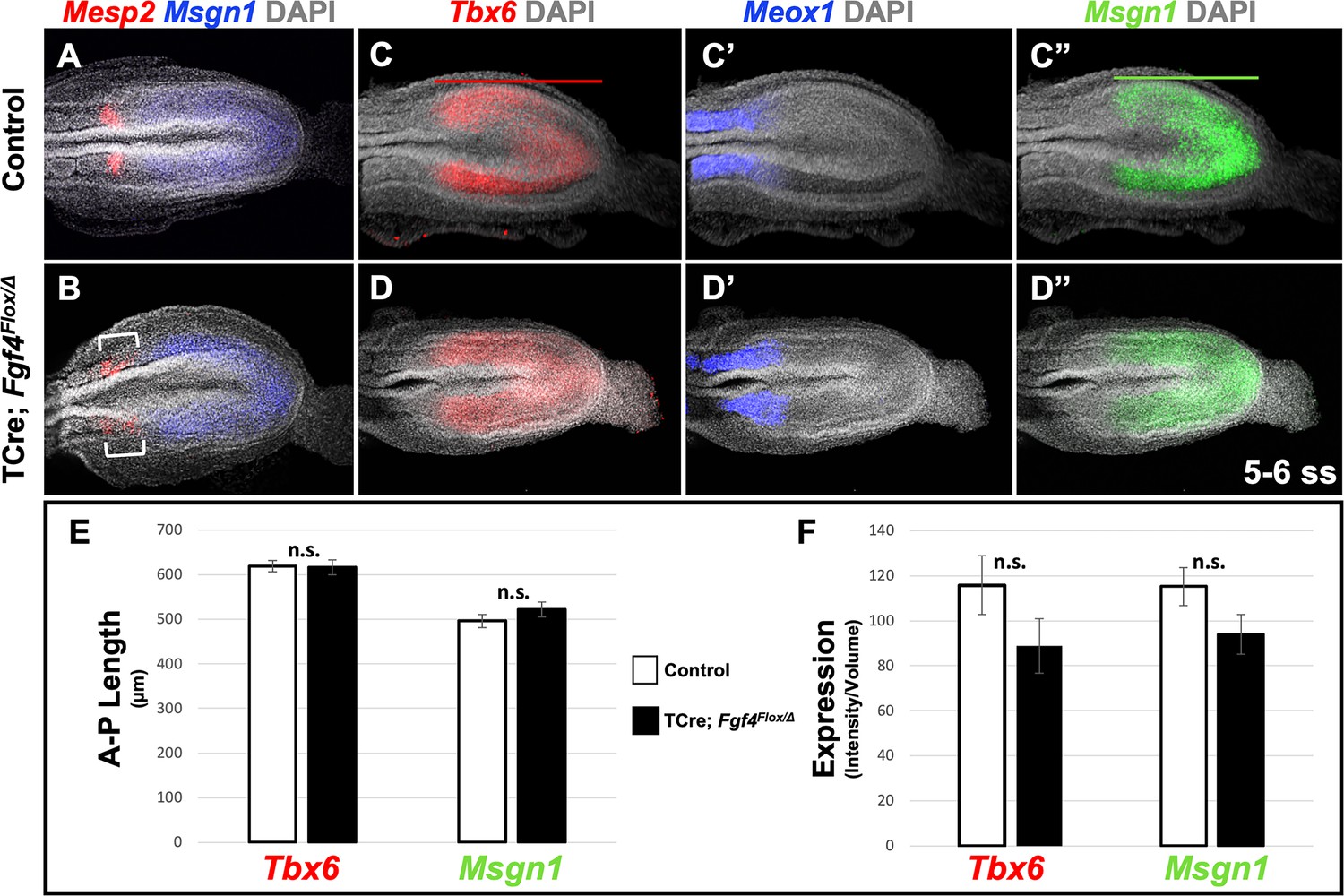
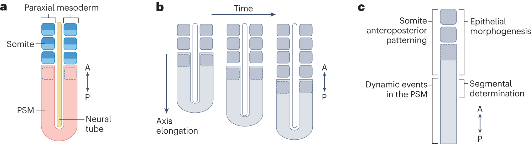
FGF (Fibroblast Growth Factor) does play a role in suppressing new somite formation, particularly in the posterior presomitic mesoderm (PSM). High levels of FGF in the posterior PSM maintain cells in an unsegmented, undifferentiated state. This suppression helps create the gradient required for the orderly, sequential formation of somites as cells move from the posterior to the anterior region of the PSM.
If FGF signaling is experimentally suppressed or reduced:
Premature Somite Formation: Cells that would normally be held in an unsegmented state in the posterior PSM can undergo premature segmentation. This can lead to the formation of somites before the cells reach the proper position in the anterior PSM, where segmentation is normally coordinated.
Disruption of Rostrocaudal Polarity: Lowered FGF levels might interfere with the normal rostrocaudal patterning within somites. FGF signaling is also involved in establishing the correct polarity within each somite, so its suppression can cause defects in somite boundary formation and polarity.
Alteration of Determination Front Position: The “determination front” — the zone where cells transition from unsegmented to segmented states — is influenced by the balance of FGF and RA gradients. Suppressing FGF shifts this determination front posteriorly, resulting in somites forming prematurely, closer to the posterior PSM.
Experimental studies have shown that reducing FGF signaling can indeed lead to the formation of smaller, irregularly patterned somites, as cells segment earlier than intended. This highlights FGF’s role in controlling the timing and spatial pattern of somite formation, ensuring that somites form in an orderly, regulated manner as cells progress through the PSM.
(ChatGPT)
FGFシグナリング、WntシグナリングによるTbx6遺伝子発現の促進
FGF and Wnt signaling are critical for activating and maintaining Tbx6 gene expression in the posterior presomitic mesoderm (PSM) during somitogenesis. Here’s how these pathways regulate Tbx6:
Activation and Maintenance of Tbx6 Expression: High levels of FGF and Wnt signaling in the posterior PSM provide a supportive environment that promotes Tbx6 expression. These signals maintain Tbx6 at levels necessary for keeping cells in an undifferentiated, mesenchymal state, preparing them for future segmentation as they move toward the anterior.
Regulation Through Gradients: FGF and Wnt form gradients that are highest in the posterior PSM and gradually decrease toward the anterior PSM. As cells “move” or are positioned closer to the anterior, they experience decreasing levels of FGF and Wnt, leading to the downregulation of Tbx6. This shift in signaling cues is essential for transitioning cells out of the Tbx6-expressing, unsegmented state, allowing them to prepare for segmentation and somite boundary formation.
Indirect Activation: While FGF and Wnt signaling are not transcription factors that bind directly to the Tbx6 promoter, they activate Tbx6 expression through downstream effectors and pathways within the cell. These downstream factors likely include various transcriptional regulators that respond to FGF and Wnt signals, which in turn promote Tbx6 transcription in the posterior PSM.
In summary, FGF and Wnt signaling indirectly activate and maintain Tbx6 expression in the posterior PSM, playing an essential role in somitogenesis by supporting Tbx6‘s role in maintaining the unsegmented, mesenchymal state of PSM cells.
(ChatGPT)
Tbx6の体節形成における抑制的な役割
In somitogenesis, Tbx6 is a key transcription factor that plays a crucial role in maintaining the undifferentiated, mesenchymal state of cells in the presomitic mesoderm (PSM) and promoting their commitment to the paraxial mesoderm lineage. This role is essential for the orderly formation of somites along the anterior-posterior axis. Here’s a detailed look at Tbx6’s role in somitogenesis:
Specifying Paraxial Mesoderm Lineage: Tbx6 helps specify cells in the PSM as paraxial mesoderm, which gives rise to somites. In the absence of Tbx6, PSM cells may adopt alternative fates, such as forming neural tissue instead of somites. This lineage specification function is critical in ensuring that the correct tissue types emerge in the right positions during development.
Maintaining Cells in an Undifferentiated State: Tbx6 expression is high in the posterior PSM, where it keeps cells in an undifferentiated and mesenchymal state, preventing them from undergoing premature segmentation. This maintenance of an undifferentiated state is essential for the proper timing of somite formation. When cells move toward the anterior PSM (注), Tbx6 expression decreases, which allows them to transition toward epithelialization and segmentation.
Coordination with FGF and Wnt Signaling: Tbx6 expression is influenced by FGF and Wnt signaling gradients, which are high in the posterior PSM and gradually decrease toward the anterior. These signaling pathways help maintain Tbx6 expression in the posterior PSM, where cells are held in a proliferative, unsegmented state. As cells approach the anterior, decreasing FGF and Wnt signals lead to downregulation of Tbx6, allowing cells to exit the proliferative state and begin the process of segmentation.
Regulation of Somite Boundary Formation: Although Tbx6 itself does not directly trigger the mesenchymal-epithelial transition (MET) needed for somite boundary formation, it creates the conditions necessary for cells to respond to other cues, such as Mesp2, that initiate boundary formation. As cells exit the Tbx6-expressing region, they become competent to form boundaries and undergo MET, marking the anterior edge of a new somite.
Interaction with Mesp2 for Rostrocaudal Polarity: Tbx6 indirectly influences the establishment of rostrocaudal (head-to-tail) polarity within somites by interacting with pathways that activate Mesp2 expression in the anterior of each forming somite. Mesp2 expression is critical for defining the anterior-posterior polarity, which is essential for the correct alignment and differentiation of somites. Proper timing of Tbx6 downregulation is necessary for Mesp2 to function appropriately in defining this polarity.
In summary, Tbx6 is essential for maintaining the mesenchymal state and paraxial mesoderm identity of cells in the PSM, coordinating with signaling pathways to control the timing and positioning of somite formation. By controlling when and where cells become competent to segment, Tbx6 ensures the sequential and ordered formation of somites, a fundamental aspect of vertebrate body segmentation.
In this context, “move toward the anterior PSM” does not imply physical movement of individual cells. Instead, it refers to the positional progression of cells along the anterior-posterior axis within the presomitic mesoderm (PSM) due to developmental processes.
More accurately, what happens is this:
Cells in the PSM are gradually positioned closer to the anterior as new cells are added posteriorly due to tail bud growth and axial elongation. This process creates a progressive shift without requiring individual cell migration.
As cells reach the anterior PSM (or the “determination front”), their exposure to signaling gradients changes (with decreased FGF/Wnt and increased RA), which influences gene expression patterns, such as the downregulation of Tbx6.
Thus, a clearer phrasing might be: “As cells are positioned closer to the anterior PSM…”
(ChatGPT)
英語のmoveという言葉には、物理的な移動だけでなく状況の変化を意味する用法もあるというわけです。
In English, the word “move” can indeed have both literal and figurative meanings, depending on the context:
Literal Meaning (Physical Movement): “Move” often implies physical movement of an object from one place to another. For example, “The car moved down the street” means the car physically traveled along the road.
Figurative or Conceptual Meaning (Change Without Physical Movement): “Move” can also describe changes that are not physical. For instance:
Position in a sequence or process: “The project moved to the next phase” implies progression in steps rather than physical movement.
Change in state or condition: “He moved to a new job role” indicates a change in status or position without physical relocation.
Shift in time or focus: “We moved the deadline” or “The story moves to a new topic” means a change in timing or subject matter, not a physical shift.
In scientific or technical language, “move” is often used figuratively to describe progression, transition, or reconfiguration within a defined space or system, even if no physical movement occurs. For instance, in biology, when we say cells “move toward differentiation,” it may mean they progress through developmental stages without physically migrating.
In somitogenesis, retinoic acid (RA) plays a key role in setting up the anterior-posterior (A-P) gradient within the presomitic mesoderm (PSM). This gradient interacts with other signaling gradients, particularly those of fibroblast growth factor (FGF) and Wnt, to establish a regulatory environment that is crucial for segmental patterning.
RA does not directly activate the expression of Mesp2, a transcription factor essential for somite boundary formation and rostrocaudal polarity. Instead, RA indirectly influences Mesp2 expression by opposing the posteriorizing effects of FGF and Wnt signaling in the PSM. High levels of FGF and Wnt signaling in the posterior PSM inhibit Mesp2 expression, whereas RA, which is synthesized in the anterior region, helps to repress FGF and Wnt as cells progress anteriorly. This repression allows the anterior PSM cells to express Mesp2 in a specific region, known as the “determination front,” where conditions are favorable for somite segmentation.
Thus, RA’s role in somitogenesis is to help balance the signaling environment in the PSM by counteracting FGF and Wnt, creating a permissive environment for Mesp2 expression at the right place and time, rather than acting as a direct activator of Mesp2.
Retinoic acid (RA) opposes FGF and Wnt signaling in the presomitic mesoderm (PSM) through several mechanisms, but it does not primarily act by directly suppressing the transcription of FGF or Wnt genes. Instead, RA counteracts FGF/Wnt signaling by influencing downstream signaling pathways and components in the following ways:
Inhibition of FGF and Wnt Signaling Pathways:
RA can indirectly interfere with FGF and Wnt signaling by modulating the expression of downstream targets or components within these pathways.
For example, RA has been shown to induce the expression of MAP kinase phosphatases (such as Dusp6), which inactivate components of the FGF signaling cascade, thereby reducing FGF signaling activity. This reduces the phosphorylation and activity of key FGF signaling molecules, effectively dampening FGF’s influence in the anterior PSM.
Antagonistic Gradient Formation:
RA and FGF/Wnt establish opposing gradients within the PSM. RA is synthesized in the anterior PSM and diffuses toward the posterior, whereas FGF and Wnt are expressed more strongly in the posterior and decrease toward the anterior.
This spatial gradient positioning allows RA to gradually counterbalance the effects of FGF and Wnt signaling as cells move anteriorly. Rather than directly suppressing FGF or Wnt gene transcription, RA establishes a microenvironment in the anterior PSM where FGF/Wnt signaling becomes progressively less dominant.
Indirect Regulation of Oscillatory and Segmental Gene Expression:
RA promotes the expression of genes required for segmentation and differentiation, such as Mesp2, which is necessary for somite boundary formation. This indirectly opposes the role of FGF and Wnt in maintaining cells in an oscillatory, undifferentiated state.
By promoting segmental gene expression, RA effectively counteracts FGF/Wnt-driven oscillatory gene activity, allowing cells to exit the oscillatory phase as they approach the determination front.
Suppression of FGF Receptor Expression:
In some studies, RA has been shown to downregulate the expression of FGF receptors (e.g., Fgfr1), which decreases the responsiveness of cells to FGF signals as they move toward the anterior PSM. This reduction in receptor expression decreases FGF pathway activity without directly impacting FGF ligand expression.
Regulation of Cell Cycle and Differentiation States:
RA promotes cell cycle exit and differentiation, which opposes the proliferative effects of FGF and Wnt signaling. By pushing cells toward a differentiated, segmentation-competent state in the anterior PSM, RA helps counteract the posteriorizing effects of FGF and Wnt, which maintain cells in a proliferative, mesenchymal, and unsegmented state.
Summary
In summary, RA opposes FGF and Wnt signaling mainly through indirect mechanisms rather than directly suppressing FGF or Wnt gene transcription. It modulates the signaling environment by affecting downstream signaling components, receptor expression, and promoting differentiation. These actions ensure that the determination front is established at the correct position within the PSM, allowing for the orderly transition from oscillatory to segmented states necessary for somite formation.
(ChatGPT 4o)
FGFやWntと周期的に変動する遺伝子との関係
In somitogenesis, FGF (Fibroblast Growth Factor) and Wnt signaling interact with Notch signaling to regulate the timing, positioning, and progression of somite formation. Here’s how FGF and Wnt influence Notch signaling within this developmental process:
1. Establishing the Posterior-to-Anterior Gradient
FGF and Wnt form a gradient across the presomitic mesoderm (PSM), with high concentrations in the posterior PSM (closer to the tail end) and decreasing toward the anterior PSM.
This gradient creates a “wavefront” that determines where cells are kept in an undifferentiated, oscillatory state versus where they are able to stop oscillating and begin somite formation.
2. Maintaining the Oscillatory State of Notch Signaling in the Posterior PSM
High FGF and Wnt levels in the posterior PSMpromote and maintain oscillatory gene expression, including that of Lfng(lunatic fringe), Hes7, and Delta in the Notch pathway.
These oscillations are crucial for the “segmentation clock” that drives periodic Notch signaling activity. FGF and Wnt signaling help sustain this clock in the posterior region by keeping cells in a proliferative, undifferentiated state.
3. Influencing the Determination Front
As cells progress anteriorly in the PSM, they encounter decreasing levels of FGF and Wnt and increasing levels of retinoic acid (RA).
At a certain threshold—called the determination front—the low levels of FGF and Wnt allow cells to exit the oscillatory Notch signaling state and stabilize their gene expression patterns, leading to somite boundary formation.
This interaction sets the position at which the oscillatory clock stops, allowing Notch signaling to stabilize and initiate differentiation.
4. Regulating Somite Boundary Formation
Notch signaling is essential for establishing the boundaries of somites, and the cessation of oscillatory Notch activity is key to determining where boundaries form.
By controlling the oscillatory vs. stabilized states of Notch signaling, FGF and Wnt ensure that somite boundaries are positioned correctly along the anterior-posterior axis.
5. Temporal and Spatial Coordination of Somite Formation
Wnt signaling also works alongside FGF to influence the periodicity of the segmentation clock by modulating the expression of clock genes like Hes7 and Lfng.
By maintaining cells in an oscillatory state until they reach the determination front, FGF and Wnt ensure that Notch signaling follows a precise temporal pattern that matches the physical positioning along the body axis, contributing to the regular, sequential formation of somites.
Summary
In somitogenesis, FGF and Wnt signaling maintain Notch signaling in an oscillatory state in the posterior PSM, essential for the segmentation clock. As cells move toward the anterior, lower FGF/Wnt levels allow them to exit this oscillatory state at the determination front, where Notch signaling stabilizes to form somite boundaries. This regulation ensures precise timing and spatial organization in somite formation.
(ChatGPT)
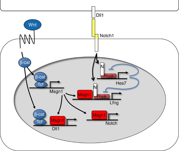
Wntの役割
下の論文のまとめの図がわかりやすいと思います。
12 July 2011 The Wnt3a/β-catenin target gene Mesogenin1 controls the segmentation clock by activating a Notch signalling program Nature Communications volume 2, Article number: 390 (2011) https://www.nature.com/articles/ncomms1381
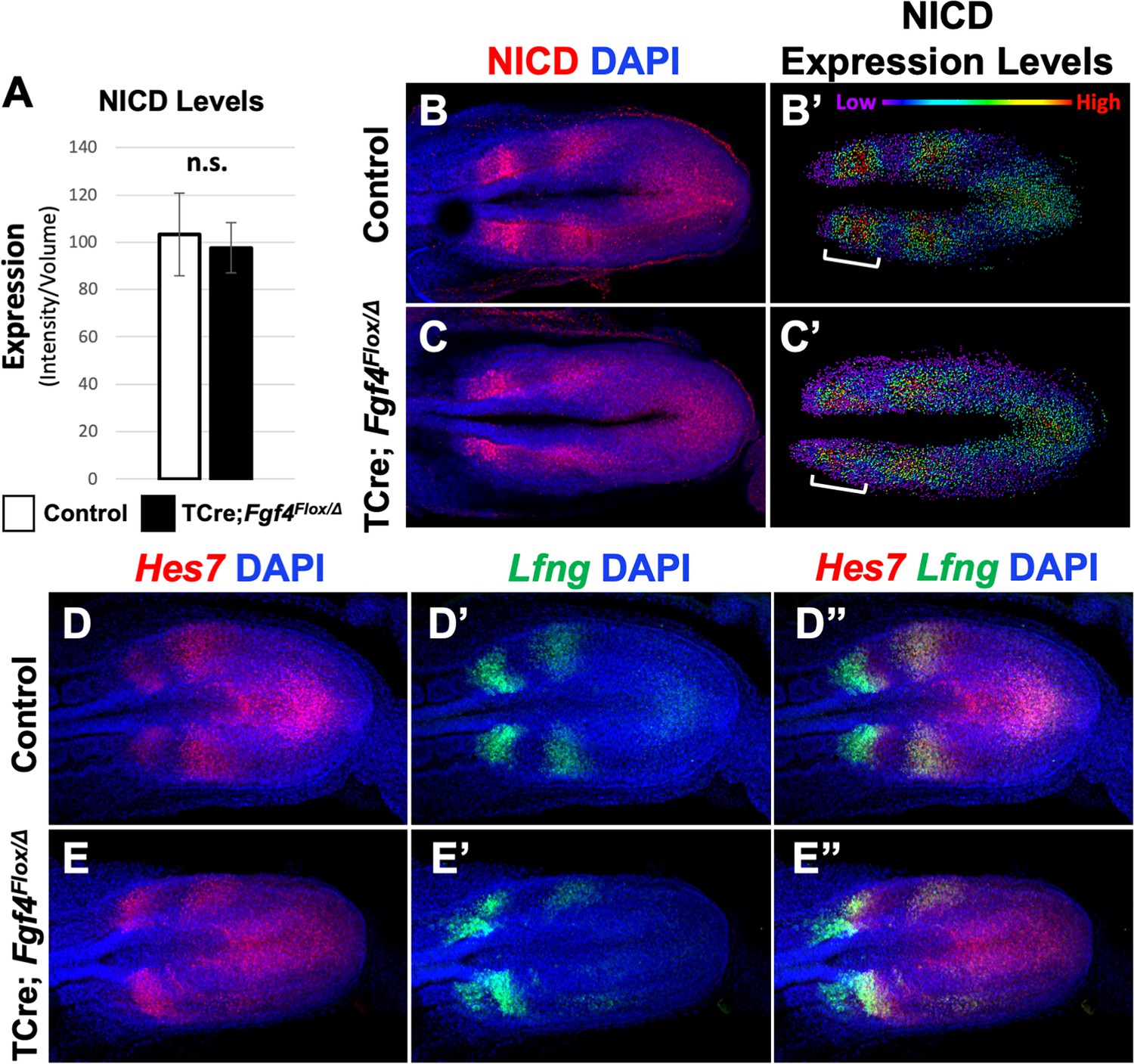
Fgf4 maintains Hes7 levels critical for normal somite segmentation clock function Nov 19, 2020 https://doi.org/10.7554/eLife.55608 https://elifesciences.org/articles/55608
シグナリング
The vertebrate Embryo Clock: Common players dancing to a different beat Front. Cell Dev. Biol., 11 August 2022 Sec. Morphogenesis and Patterning Volume 10 – 2022 | https://doi.org/10.3389/fcell.2022.944016
In the oscillatory system of somitogenesis, Notch signaling is generally considered the most upstream component that initiates and regulates the segmentation clock, of which HES genes (like Hes1 and Hes7) are key downstream oscillatory elements.
Reasons Why Notch is Upstream of HES
Activation of HES Genes by Notch Signaling:
Notch signaling is activated when Delta ligands bind to Notch receptors on neighboring cells. This interaction triggers the Notch intracellular domain (NICD) to be released and translocate to the nucleus, where it activates the transcription of downstream target genes, including the HES family (e.g., Hes1, Hes7).
Thus, Notch signaling initiates the oscillatory expression of HES genes in the presomitic mesoderm (PSM).
Synchronization Role of Notch:
Notch signaling is also critical for synchronizing oscillations between neighboring cells in the PSM, ensuring coordinated oscillatory gene expression across cells that will form a somite boundary. This intercellular communication mediated by Notch and Delta helps maintain the rhythmic, synchronized oscillations of HES genes across the tissue.
Regulation by FGF and Wnt Gradients:
Notch activity, influenced by upstream FGF and Wnt gradients, helps establish the oscillatory environment in the posterior PSM. These gradients set up the context for Notch signaling to initiate the oscillations, positioning Notch as a primary regulator in the segmentation clock.
Feedback Mechanism with HES Genes:
HES proteins, once expressed, participate in a negative feedback loop to repress their own transcription and the transcription of other oscillatory genes. However, without initial Notch signaling, this feedback loop in HES genes would not be activated, making Notch the upstream trigger.
Summary
In somitogenesis, Notch signaling is the most upstream component of the oscillatory system, activating and coordinating the expression of HES genes and other oscillatory genes within the segmentation clock. HES genes then function in feedback loops as downstream effectors, creating the cyclic gene expression patterns essential for somite formation.
Notchは周期的に変動しないことに関して
In somitogenesis, Notch gene expression itself does not oscillate like Delta; rather, Notch signaling activity oscillates due to the oscillatory expression of its ligands (like Delta) and downstream effectors (such as HES genes).
Here’s how Notch signaling oscillations occur without Notch gene expression itself being oscillatory:
Constant Notch Receptor Expression:
The expression of Notch receptors (such as Notch1 in mice) remains relatively stable in the presomitic mesoderm (PSM) and does not oscillate. Instead, the Notch receptor is always present on the cell surface, ready to respond to Delta ligand binding.
Oscillatory Delta Ligand Expression:
Delta ligands (e.g., Dll1 in mice) do oscillate in their expression, driven by the segmentation clock. When Delta levels rise on the surface of one cell, it binds to Notch receptors on neighboring cells, activating Notch signaling in those cells in an oscillatory manner.
This oscillatory expression of Delta ligand initiates periodic activation of Notch signaling in adjacent cells, creating a synchronized oscillation of Notch pathway activity across the PSM.
Oscillatory Downstream Targets (HES Genes):
The activation of Notch by oscillating Delta triggers the expression of downstream targets like HES genes (e.g., Hes1, Hes7), which also oscillate. HES proteins provide negative feedback that helps control the oscillatory cycle of Delta and other segmentation clock genes.
Intercellular Synchronization:
The stable presence of Notch receptors and the oscillatory expression of Delta enable the synchronized activation of Notch signaling across neighboring cells. This intercellular communication is critical for maintaining coordinated oscillations in the PSM, which ultimately determine the timing and location of somite boundaries.
Summary
While Notch gene expression itself does not oscillate in somitogenesis, the Notch signaling pathway oscillates due to the periodic expression of Delta ligands and HES gene feedback loops. This oscillatory signaling is essential for the segmentation clock and synchronized somite formation across the presomitic mesoderm.
Notch-Deltaシグナリングの役割
Notch Signaling Pathway | Purpose and Mechanism JJ Medicine チャンネル登録者数 107万人
デルタは遺伝子発現の変動が知られています。
Oscillatory control of Delta-like1 in cell interactions regulates dynamic gene expression and tissue morphogenesis Genes & Dev. 2016. 30: 102-116 https://genesdev.cshlp.org/content/30/1/102/F1.expansion.html
10 May 2024 Oscillatory control of embryonic development Development (2024) 151 (9): dev202191. https://journals.biologists.com/dev/article/151/9/dev202191/347230/Oscillatory-control-of-embryonic-development
Pulses of Notch activation synchronise oscillating somite cells and entrain the zebrafish segmentation clock Cristian Soza-Ried, Emre Öztürk, David Ish-Horowicz, Julian Lewis Author and article information Development (2014) 141 (8): 1780–1788. 15 April 2014 https://doi.org/10.1242/dev.102111
Notch intracellular domain (NICD), which translocates to the nucleus and associates with the DNA binding protein CSL [CBF1/RBPjκ/Su(H)/Lag1] to activate gene expression. luciferase complementation imaging (LCI) reporters for NICD-CSL association https://pubmed.ncbi.nlm.nih.gov/21775282/
Oscillatory Notch signalling during somite boundary formation, imaged using SPIM The Company of Biologists チャンネル登録者数 5930人
同様にChatGPTの回答です。
In somitogenesis, the Notch-Delta signaling pathway is essential for coordinating the timing and spatial organization of somite formation. This pathway functions as part of a molecular “segmentation clock,” which ensures that somites form in a regular, sequential manner along the anterior-posterior (A-P) axis of the developing embryo.
Key roles of Notch-Delta signaling in somitogenesis include:
Oscillatory Gene Expression: Notch-Delta signaling drives oscillations of specific “clock genes” in the presomitic mesoderm (PSM), such as Hairy1, Hes7, and others, which cycle in a periodic manner. This oscillation occurs with a set frequency, creating a wavefront of gene expression that moves through the PSM. This “segmentation clock” is crucial for determining the periodicity of somite formation.
Synchronization of Cells: Notch-Delta signaling helps synchronize the oscillatory behavior of neighboring cells in the PSM. Through intercellular communication, Delta ligands from one cell bind to Notch receptors on adjacent cells, coordinating their segmentation clocks. This synchronization ensures that cells within a future somite boundary express genes likeMesp2 together, enabling the precise formation of somites.
Boundary Formation and Rostrocaudal Polarity: Notch signaling is critical in specifying somite boundaries and establishing rostrocaudal (head-to-tail) polarity within each somite. At the determination front, where somites bud off from the PSM, Notch signaling interacts with factors like Mesp2 to trigger the epithelialization process that creates the somite boundary. The periodic activation of Notch also helps establish differential gene expression within each somite, giving them a distinct anterior and posterior polarity.
Coordination with FGF and RA Gradients: Notch-Delta signaling is integrated with FGF and RA gradients in the PSM, which help establish the anterior-posterior axis. Notch oscillations occur within a field shaped by these gradients, defining the region where somites form (the “determination front”). As the PSM cells move out of the FGF-rich posterior region toward the RA-rich anterior region, Notch signaling cues become crucial in determining where and when a somite boundary will form.
In summary, Notch-Delta signaling coordinates the temporal and spatial patterning of somite formation by driving oscillatory gene expression, synchronizing cell behaviors, defining boundaries, and interacting with other signaling pathways. This fine-tuned regulation is critical for forming the segmented structure of the vertebrate body.
(ChatGPT)
In somitogenesis, Delta (a ligand in the Notch signaling pathway) exhibits oscillatory gene expression primarily through its regulation by the segmentation clock, which is driven by negative feedback loops involving oscillatory genes like Hes family members (e.g., Hes1 and Hes7). Here’s a breakdown of the mechanism behind Delta’s oscillatory expression:
Notch-Delta Signaling and Feedback Loop:
Delta ligands (e.g., Dll1 in mice) are part of the Notch signaling pathway, which is crucial for coordinating the segmentation clock across neighboring cells in the presomitic mesoderm (PSM).
When Delta binds to Notch receptors on neighboring cells, it activates the Notch pathway, leading to the expression of downstream genes, including Hes genes. This interaction synchronizes oscillatory gene expression between neighboring cells, which is essential for organized somite formation.
Hes Genes as Transcriptional Repressors:
Hes genes, activated by Notch signaling, function as transcriptional repressors and play a key role in generating oscillations. For example, Hes7represses its own transcription as well as that of Delta by binding to their promoters, creating a negative feedback loop.
After Hes proteins are synthesized, they inhibit further transcription of their own gene and Delta. As Hes proteins degrade, repression is lifted, allowing Delta and Hes transcription to restart, resulting in a cycle of oscillatory expression.
Negative Feedback with a Time Delay:
This oscillatory behavior relies on a time delay in the negative feedback loop, which is a function of mRNA and protein synthesis, degradation rates, and cellular delays in transcriptional repression.
The degradation of Hes proteins and mRNA is relatively rapid, and once they are sufficiently degraded, repression is released, allowing a new cycle of Delta and Hes expression to begin. This delay-dependent negative feedback is critical for generating stable oscillations.
Synchronization Across Cells via Notch-Delta Interactions:
Notch-Delta signaling between neighboring cells helps synchronize their oscillatory patterns. Delta expression in one cell activates Notch in an adjacent cell, which in turn influences Hes gene expression in that neighboring cell, creating coordinated oscillations.
This intercellular coupling ensures that Delta expression oscillates synchronously across cells in the PSM, which is essential for creating uniform, periodic somite boundaries.
Interaction with FGF and Wnt Gradients:
Delta oscillations also interact with the FGF and Wnt signaling gradients across the PSM. These gradients are high in the posterior PSM and decrease toward the anterior, modulating the pace and location of Delta oscillations.
As cells move toward the anterior PSM, where FGF and Wnt levels are lower, Delta oscillations slow and eventually stabilize, marking the transition point where somite boundaries begin to form.
Summary
Delta’s oscillatory expression in somitogenesis is driven by a negative feedback loop involving Hes genes, with a time delay that produces cyclical repression and derepression. This oscillation is synchronized across cells by Notch-Delta signaling between neighboring cells, and modulated by the FGF and Wnt gradients to ensure coordinated somite formation.
Lunatic fringe (Lfng) gene expression oscillates in somitogenesis, and it is driven by Notch signaling. Here’s a detailed look at why and how this oscillation occurs:
1. Role of Notch Signaling in Driving Lunatic Fringe Oscillations
Notch Activation: The oscillatory expression of Lfng is a result of periodic Notch signaling activation in the presomitic mesoderm (PSM). Notch signaling is activated by ligands such as Delta-like 1 (Dll1), which bind to Notch receptors on neighboring cells.
Transcriptional Activation of Lfng: When Notch receptors are activated, they release the Notch intracellular domain (NICD), which translocates to the nucleus and activates transcription of target genes, including Lfng.
Negative Feedback and Oscillations: Lfng itself plays a role in modulating Notch signaling by glycosylating the Notch receptor, which modifies its responsiveness to Delta ligands. This feedback can suppress further activation of Notch in a periodic manner. This creates a negative feedback loop where Lfng expression periodically increases and decreases, creating oscillations.
2. Why Lunatic Fringe Needs to Oscillate
Coordination with the Segmentation Clock: Oscillatory expression of Lfng is synchronized with other segmentation clock genes (such as Hes7), ensuring that Notch signaling oscillates in a coordinated manner across the PSM. This synchronization is critical for the precise timing of somite boundary formation.
Control of Somite Boundary Formation: Lfng oscillations help regulate when and where Notch signaling is active along the anterior-posterior axis, contributing to the orderly segmentation needed for somite boundary formation.
3. Interaction with Other Signals
Wnt and FGF Gradients: While Notch signaling directly drives Lfng oscillations, the activity of Notch signaling itself is modulated by Wnt and FGF gradients across the PSM. High levels of Wnt and FGF in the posterior PSM sustain the oscillatory environment, maintaining Notch signaling activity. As cells move toward the anterior and encounter lower levels of Wnt and FGF, they begin to stabilize their gene expression patterns, including Lfng, as they prepare for somite formation.
Negative Feedback in the Notch Pathway: In addition to Lfng, other Notch pathway components like Hes genes also oscillate and contribute to the timing and coordination of Notch-driven processes.
Summary
Notch signaling drives the oscillatory expression of Lunatic fringe (Lfng) in somitogenesis. The oscillations in Lfng are a result of Notch activation by ligands like Delta, combined with a negative feedback loop where Lfng itself modulates Notch receptor sensitivity. This oscillation is synchronized with other segmentation clock genes, allowing precise timing and spatial organization for somite boundary formation. The stability of Lfng oscillations depends on Notch signaling, which is further modulated by posterior Wnt and FGF gradients.
(ChatGTP 4o)
Lunatic fringe (often abbreviated as Lfng) is a gene that encodes an enzyme known as a glycosyltransferase, which modifies the Notch receptor, influencing how it responds to Delta and other Notch ligands. It plays an important role in regulating Notch signaling oscillations during somitogenesis. Here’s a breakdown of lunatic fringe’s function, its name, and how it fits into the Notch signaling pathway.
1. What Lunatic Fringe Does
Role in Notch Signaling: Lunatic fringe modifies the Notch receptor by adding sugars to it (glycosylation). This modification changes the sensitivity of Notch to its ligands, including Delta-like (Dll1) and Jagged ligands.
Oscillatory Expression: Like Delta and HES genes, Lfng expression oscillates in the presomitic mesoderm (PSM) as part of the segmentation clock. These oscillations help regulate the timing of Notch activation, contributing to the rhythmic formation of somites.
Function in Somite Formation: By modulating Notch signaling, lunatic fringe helps maintain the periodic activation of Notch in a way that coordinates somite segmentation across neighboring cells.
2. Literal Meaning of the Name “Lunatic Fringe”
The name “lunatic fringe” is somewhat playful, inspired by the idea of an “edge” or “fringe” that behaves in a periodic or “lunatic” (crazy, unpredictable) manner.
Fringe refers to the family of fringe proteins (other members include manic fringe and radical fringe), which all influence Notch signaling by modifying the Notch receptor’s response to its ligands.
Lunatic implies something erratic or periodic, likely a nod to the cyclic or “oscillatory” behavior seen in Notch signaling and segmentation, somewhat like the moon’s periodic phases (the root “luna” refers to the moon).
Summary
Lunatic fringe is a glycosyltransferase enzyme that modifies the Notch receptor, affecting how it responds to ligands like Delta. Its oscillatory expression is crucial for somite formation in somitogenesis. While it is neither Delta nor HES, it plays a critical role in controlling the oscillations of Notch signaling that drive the segmentation clock. The name “lunatic fringe” reflects the periodic or “fringe” nature of its action in the oscillatory system of somitogenesis.
Imaging Clock Oscillations in the Mouse Embryo Cell Press チャンネル登録者数 2.58万人
Wave of gene expression in an embryo European Molecular Biology Laboratory (EMBL) チャンネル登録者数 1.34万人 In vivo imaging of gene activity oscillations — Two-photon time-lapse imaging of using the LuVeLu transgenic line (reporting lunatic fringe gene activity); side view on the paraxial mesoderm, merged YFP and brightfield channel. Real-time recordings reveal oscillatory patterns of gene activity (Venus channel), sweeping from posterior to anterior mesoderm as periodic waves. Simultaneously, segment formation occurs where waves come to halt. Credit: Nature
Travelling waves in somitogenesis: collective cellular properties emerge from time-delayed juxtacrine oscillation coupling Progress in Biophysics and Molecular Biology doi: 10.1016/j.pbiomolbio.2018.04.004 https://linkinghub.elsevier.com/retrieve/pii/S0079-6107(18)30017-8 本文有料 https://www.biorxiv.org/content/10.1101/297671v1.full
Hairy/HES1
HES1はHairy and Enhancer of Split 1の略で、Hairy遺伝子ファミリーの一部です。 HES1は、哺乳類のHairyホモログであり、Notchシグナル伝達経路によって誘導される転写因子です。もともとHairyはショウジョウバエ(Drosophila)で発見され、発生中の体節形成や遺伝子発現の制御に関与しています。哺乳類では、HES1やHES7などが体節形成や神経発生の際に遺伝子発現の振動を担い、体の発生において重要な役割を果たしています。 このため、HES1は「哺乳類のHairy遺伝子の一種」として位置づけられ、振動的な発現パターンを示す遺伝子として、発生学的な研究で頻繁に参照される遺伝子の1つです。
Avian hairy Gene Expression Identifies a Molecular Clock Linked to Vertebrate Segmentation and Somitogenesis Isabel Palmeirim1 ∙ Domingos Henrique2,§ ∙ David Ish-Horowicz2 ∙ Olivier Pourquié CELL Volume 91, Issue 5p639-648November 28, 1997 https://www.cell.com/fulltext/S0092-8674%2800%2980451-1
Autoinhibition with Transcriptional Delay: A Simple Mechanism for the Zebrafish Somitogenesis Oscillator Julian Lewis Current Biology Volume 13, Issue 16, 19 August 2003, Pages 1398-1408 Journal home page for Current Biology Article https://doi.org/10.1016/S0960-9822(03)00534-7 https://www.sciencedirect.com/science/article/pii/S0960982203005347
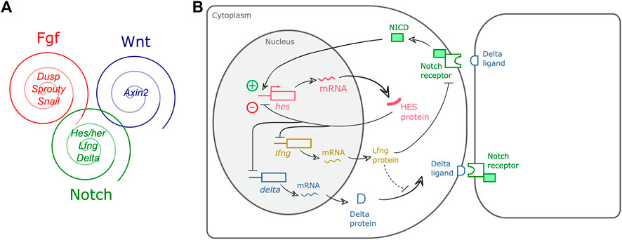
In somitogenesis, Notch, WNT, and FGF signaling pathways all play critical roles, but their oscillatory behaviors vary:
1. Notch Pathway
Oscillatory Behavior: YES
Genes in the Notch signaling pathway, such as Hes/Hairy family transcription factors, exhibit robust oscillatory expression in the presomitic mesoderm (PSM).
Oscillations are driven by transcriptional feedback loops:
Hes1 represses its own transcription, leading to periodic expression cycles.
These oscillations are synchronized among neighboring cells through Notch-mediated intercellular communication.
2. WNT Pathway
Oscillatory Behavior: YES
Components of the WNT signaling pathway, like Axin2, exhibit oscillatory expression.
Oscillations in the WNT pathway are driven by negative feedback loops involving β-catenin stabilization and Axin2 expression.
These oscillations are slower compared to Notch oscillations and are thought to interact with both Notch and FGF pathways.
3. FGF Pathway
Oscillatory Behavior: CONTROVERSIAL / INDIRECT
FGF signaling itself is not directly oscillatory, but its downstream targets, such as Dusp6 and Spry4, show oscillatory expression in some species (e.g., zebrafish).
FGF’s gradient along the PSM establishes a “wavefront” rather than an intrinsic oscillator. However, this wavefront interacts with the oscillatory clock (Notch and WNT) to define the periodic segmentation of somites.
Summary of Oscillatory Behavior
Pathway
Oscillatory Expression
Key Oscillatory Genes
Notch
Yes
Hes1, Hes7, Lfng
WNT
Yes
Axin2, Dkk1
FGF
Indirect/Controversial
Dusp6, Spry4 (in zebrafish and some species)
Interaction Between Pathways
Coupling of Oscillators:
Notch, WNT, and (indirectly) FGF oscillators are coupled to ensure coordinated somite formation.
Wavefront Mechanism:
The FGF signaling gradient works as a “wavefront” that interacts with the Notch and WNT “clock” to regulate the timing and position of somite segmentation.
In conclusion, Notch and WNT pathways show clear oscillatory gene expression, while FGF signaling oscillations are species-dependent and often secondary to other oscillatory systems. Together, these pathways synchronize to drive the somitogenesis clock.
The FGF signaling gradient interacts with the oscillatory clock genes (primarily from the Notch and WNT pathways) to coordinate the segmentation process during somitogenesis. Here’s a detailed explanation of this interaction:
1. Overview of the FGF Gradient
Gradient Formation:
The FGF signaling pathway establishes a posterior-to-anterior gradient in the presomitic mesoderm (PSM).
FGF8 (and to some extent FGF4) is produced in the posterior PSM and diffuses anteriorly, creating a concentration gradient.
Degradation of FGF mRNA (e.g., by retinoic acid signaling from the anterior) ensures a sharp decline in FGF signaling towards the anterior.
Function:
The FGF gradient defines a wavefront, also known as the “determination front,” which interacts with the oscillatory clock genes to determine the spatial boundary for somite formation.
2. Oscillatory Clock Genes
Genes from the Notch (e.g., Hes7) and WNT (e.g., Axin2) pathways oscillate periodically in the PSM.
These oscillations are synchronized within and between cells, creating waves of gene expression that travel posterior-to-anterior in the PSM.
3. Interaction Between FGF Gradient and Clock Genes
The FGF gradient interacts with the clock genes in a spatiotemporal manner to regulate somite segmentation:
(1) Threshold Mechanism
Anterior FGF Threshold:
Cells in the PSM “read” the FGF gradient. When the FGF concentration drops below a certain threshold, oscillatory clock gene expression stops, and the cells are primed to form a somite.
This threshold defines the anterior boundary of a new somite.
Coupling with Oscillations:
The clock genes, which oscillate in a periodic manner, are interpreted by the wavefront (set by the FGF gradient) to determine when and where segmentation occurs.
(2) Synchronization of Timing
The oscillatory cycles of clock genes determine the timing of somite formation.
The FGF gradient sets the spatial position where the next somite boundary will form.
(3) WNT-FGF Feedback Loop
WNT-Dependent Regulation of FGF:
WNT signaling in the posterior PSM maintains FGF expression. This ensures the FGF gradient remains sharp and dynamic.
FGF’s Effect on WNT:
FGF signaling can modulate the WNT pathway through feedback loops, ensuring that WNT-driven oscillations are synchronized with the FGF gradient.
4. Molecular Interactions
FGF and Clock Gene Regulation:
FGF Downstream Genes:
Dusp6, Spry2, and Spry4 are feedback regulators of FGF signaling. These genes oscillate in the posterior PSM in some species (e.g., zebrafish), indirectly linking FGF signaling to the oscillatory clock.
Clock Gene Impact on FGF Gradient:
The segmentation clock genes regulate the Mesp2 expression at the wavefront, which is critical for determining somite boundaries. Mesp2 expression is tightly regulated by the interplay between the FGF gradient and Notch signaling.
5. Spatiotemporal Dynamics
The interaction of the FGF gradient and the oscillatory clock genes ensures that:
Timing:
The oscillatory clock sets the periodicity of somite formation (e.g., ~90 minutes in mice).
Position:
The FGF gradient sets the spatial location where somite boundaries form.
Coordination:
As the FGF gradient moves posteriorly due to axial elongation, it provides a moving reference for the oscillations to interact with.
6. Evolutionary and Experimental Insights
Species-Specific Differences:
In zebrafish, FGF signaling shows oscillatory expression (e.g., Dusp6), whereas in mice, FGF is primarily a static gradient with a wavefront function.
Perturbation Studies:
Disrupting FGF signaling (e.g., by inhibiting FGF receptors) abolishes the ability of the wavefront to interact with oscillatory clock genes, leading to defective somite formation.
7. Summary
The FGF gradient serves as a wavefront that interacts with oscillatory clock genes to spatially and temporally coordinate somite formation.
Key mechanisms:
FGF concentration below a threshold stops oscillations and triggers somite segmentation.
The interplay between FGF and WNT signaling ensures proper maintenance of the gradient and its alignment with clock oscillations.
This dynamic interaction ensures the periodic formation of somites, which is critical for proper vertebrate body segmentation.
This interaction showcases a remarkable example of how temporal oscillations and spatial gradients integrate to create precise developmental patterns.
FGFの濃度が下がるとなぜ振動が止まるのでしょうか。これもChatGPTに訊いてみます。
The drop in FGF concentration triggers the cessation of oscillations in the presomitic mesoderm (PSM) because the FGF signaling pathway regulates the transcriptional and translational dynamics necessary for maintaining the oscillatory clock. The mechanisms underlying this process involve threshold regulation, cross-talk with WNT and Notch pathways, and transcriptional control of critical genes. Below is a detailed explanation:
1. Role of FGF in Maintaining Oscillations
FGF signaling sustains the expression of oscillatory clock genes (e.g., Hes7 in the Notch pathway and Axin2 in the WNT pathway) in the posterior PSM.
High FGF concentration in the posterior:
Promotes active transcription of clock genes.
Supports the dynamic feedback loops necessary for oscillations.
As cells move anteriorly in the PSM, they experience progressively lower FGF concentrations due to the FGF gradient.
2. Mechanisms for Oscillation Cessation
(1) Threshold Mechanism
FGF concentration-dependent transition:
FGF levels drop below a threshold as cells migrate anteriorly.
This low FGF signaling environment disrupts the transcriptional feedback loops that drive oscillations.
(2) FGF’s Regulation of Clock Genes
Notch Pathway Genes:
FGF signaling supports the periodic expression of Hes7 by regulating upstream Notch pathway components.
Loss of FGF signaling destabilizes Hes7’s negative feedback loop, halting oscillations.
WNT Pathway Genes:
FGF maintains WNT signaling in the posterior PSM, ensuring oscillatory behavior of genes like Axin2.
A reduction in FGF signaling decreases WNT activity, which is critical for oscillation maintenance.
(3) FGF-Dependent Transcriptional Control
Mesp2 Activation:
When FGF concentration drops below the threshold, the transcription factor Mesp2 is upregulatedin the anterior PSM.
Mesp2 represses genes involved in the clock’s oscillatory machinery (e.g., Hes7), promoting stabilization rather than continued oscillation.
FGF Suppresses Mesp2:
High FGF levels inhibit Mesp2 expression in the posterior PSM, keeping cells in an oscillatory state.
Loss of this suppression allows Mesp2 to act, halting oscillations and initiating somite boundary formation.
(4) Cross-Talk with Retinoic Acid (RA)
FGF and RA gradients are antagonistic:
Retinoic acid (RA), which is concentrated in the anterior PSM, counteracts FGF signaling.
As cells move anteriorly and experience lower FGF levels, RA activity increases, further suppressing clock gene oscillations.
RA promotes Mesp2 expression, solidifying the cessation of oscillations.
(5) Epigenetic and Translational Changes
FGF signaling maintains the permissive transcriptional and translational environment necessary for oscillations by:
Regulating the activity of transcription factors like Tbx6, which is essential for maintaining the oscillatory state.
Supporting ribosomal function and RNA processing for rapid feedback regulation.
A drop in FGF signaling results in transcriptional repression and slowed feedback dynamics, halting oscillatory behavior.
3. Experimental Evidence
FGF Inhibition Studies:
Pharmacological inhibitors of FGF signaling (e.g., SU5402) in mouse and zebrafish embryos disrupt oscillations in the PSM and prematurely stabilize the expression of clock genes.
FGF-RA Interaction Studies:
Altering the balance of FGF and RA gradients shifts the location of oscillation cessation, confirming their antagonistic role in regulating the clock.
Genetic Models:
Knockout models of FGF pathway components (e.g., Fgf8 or Sprouty genes) show defective segmentation and premature cessation of oscillations.
4. Conceptual Summary
The FGF gradient functions as a spatial timer:
In regions of high FGF, oscillations are active due to the maintenance of feedback loops.
In regions of low FGF, feedback loops are destabilized, leading to oscillation cessation.
The cessation of oscillations is coupled with:
Mesp2 activation.
Transition to a stabilized gene expression state necessary for somite boundary formation.
This mechanism ensures that the oscillatory “clock” and the spatial “wavefront” are tightly coordinated, allowing for precise somite segmentation.
モルフォゲンと振動で次の体節がどう決まるのか
How Does the Gradient Interact with the Segmentation Clock? Signaling Gradients during Paraxial Mesoderm Development Alexander Aulehla and Olivier Pourquie Cold Spring Harb Perspect Biol 2010;2:a000869 https://cshperspectives.cshlp.org/content/2/2/a000869.full
新しく形成される体節の「境界」を決めるシグナル
ChatGPTの回答。
The specification of the caudal (posterior) boundary of a newly formed somite is tightly regulated by several interacting signaling pathways and genes. Key signals and regulatory elements involved include:
Notch Signaling: Notch signaling is crucial for determining the rostrocaudal (head-to-tail) polarity within each somite. In particular, Mesp2, a downstream target of Notch, is expressed in the anterior portion of the forming somite. This differential expression contributes to defining the boundary between the rostral (anterior) and caudal (posterior) compartments of each somite. Notch activity helps restrict Mesp2 expression, thereby influencing the formation of the boundary at the caudal end.
FGF and Wnt Gradients: Fibroblast Growth Factor (FGF) and Wnt signaling gradients are high in the posterior presomitic mesoderm (PSM) and gradually decrease toward the anterior. As cells move anteriorly within the PSM, they leave the influence of high FGF and Wnt levels, which allows for changes in gene expression critical for somite boundary formation. These gradients work together with Notch signaling to ensure that cells in the anterior PSM can form somites with distinct anterior and posterior regions.
RA (Retinoic Acid) Gradient: Retinoic acid, produced in the anterior PSM, creates a gradient opposing that of FGF and Wnt. This gradient helps create a permissive environment in the anterior PSM for somite boundary formation. The balance between RA and FGF/Wnt gradients helps set up the “determination front” where somites form, with the anterior boundary of the new somite influenced by RA and the posterior boundary still affected by FGF and Wnt signaling.
Tbx6 and Ripply Genes: Tbx6, a transcription factor expressed in the posterior PSM, helps maintain the unsegmented state of cells in this region. As cells reach the determination front, Tbx6 expression is downregulated in a process partly mediated by the Ripply genes, which are also regulated by Mesp2. This downregulation allows for the differentiation of the anterior and posterior compartments of the somite, specifying the caudal boundary of the newly formed somite.
Ephrin/Eph Signaling: Once the somite boundary has been established, Ephrin/Eph signaling becomes active at the newly formed boundary, reinforcing segmentation and boundary stabilization. Ephrin ligands are typically expressed in the posterior cells, while Eph receptors are in the anterior cells of the next forming somite. This interaction helps create a clear caudal boundary for each somite.
Together, these signals and gene interactions establish the caudal boundary of the somite, ensure correct rostrocaudal polarity, and contribute to the distinct segmentation pattern necessary for organized vertebrate body plan development.
When a new somite forms, the establishment of its anterior and posterior boundaries is a successive process rather than a simultaneous one.
Here’s a breakdown of how these boundaries form:
Posterior Boundary Formation: The posterior boundary of the new somite is defined first. This boundary is established as cells in the presomitic mesoderm (PSM) reach the “determination front,” where they encounter conditions favorable for somite segmentation. At this front, signals such as reduced FGF and Wnt levels (alongside elevated RA levels in the anterior PSM) allow these cells to undergo the segmentation process. Notch signaling also plays a crucial role here, helping to define the initial boundary.
Anterior Boundary Formation: After the posterior boundary is defined, the anterior boundary of the forming somite is established. This is typically marked by the differential expression of genes like Mesp2, which is initially restricted to the rostral (anterior) half of the somite. This expression leads to the establishment of the anterior boundary, defining the complete somite with distinct anterior and posterior halves.
This stepwise process ensures that each somite forms with the correct orientation and rostrocaudal polarity. By establishing boundaries sequentially, the somitogenesis machinery ensures that each somite is properly aligned with its neighboring segments, creating the precise, segmented structure essential for vertebrate body patterning.
(ChatGPT)
間葉-上皮 転換
In somitogenesis, the Mesenchymal-Epithelial Transition (MET), which is essential for somite boundary formation, involves the gene Paraxis as well as other key players. Here’s how these genes contribute to the MET process:
Paraxis: Paraxis is a basic helix-loop-helix (bHLH) transcription factor that is crucial for the epithelialization of somites. Paraxis promotes the transition of mesenchymal cells in the presomitic mesoderm (PSM) to a more epithelial, organized structure within each somite. In Paraxis mutants, somites fail to undergo proper epithelialization, resulting in disorganized, mesenchymal-like structures rather than well-defined somites.
E-cadherin and N-cadherin: Cadherins are adhesion molecules essential for establishing and maintaining the epithelial structure of cells. During MET, cells in the forming somite upregulate cadherins (especially N-cadherin), promoting cell-cell adhesion and the formation of a segmented epithelial structure.
Fibronectin and Integrins: These extracellular matrix (ECM) components and receptors help stabilize the epithelial structure within somites. Fibronectin provides structural support in the ECM, and integrins on the cell surface interact with fibronectin to promote cell adhesion and polarity, reinforcing the MET process.
Notch Signaling and Mesp2: Notch signaling, via Mesp2, also influences the MET process. Mesp2 expression in the anterior portion of each forming somite contributes to boundary formation and epithelialization, working in concert with Paraxis and cadherins to establish the segmented, epithelial state.
Together, these genes and molecular pathways drive the mesenchymal-epithelial transition in somitogenesis, enabling the formation of organized, epithelialized somites essential for the segmented structure of the vertebrate body plan.
(ChatGPT)
上の説明では体節の前半分という話がでてきましたが、後ろ半分に関してはどうなのでしょうか。
In the posterior portion of each forming somite, the mesenchymal-epithelial transition (MET) and establishment of somite boundaries are regulated by specific genes that contribute to the segmentation and polarization of somites. Key genes essential for MET and somite formation in the posterior region include:
Mesp2: Mesp2 is a crucial transcription factor that plays a role in defining the anterior-posterior polarity within each somite. In the anterior portion of a forming somite, Mesp2 expression helps initiate MET and boundary formation. This, in turn, indirectly defines the posterior boundary by establishing a sharp contrast with the posterior region, which has lower Mesp2 expression.
Notch Signaling Pathway: The Notch pathway is integral to maintaining the segmentation clock and ensuring that the posterior PSM cells enter a synchronized cycle of differentiation and segmentation. Notch signaling also helps specify the rostrocaudal polarity within each somite. In particular, genes such as Dll1 (Delta-like 1) and Lfng (Lunatic fringe) within the Notch pathway show oscillatory expression patterns that help set the timing and boundary for somite formation. Notch activity is crucial for the posterior portion to remain distinct from the anterior and to respond appropriately during the MET.
Uncx4.1 (Uncx): Uncx4.1 is expressed specifically in the posterior half of each somite after it forms. This transcription factor helps maintain the identity and structure of the posterior compartment and is essential for establishing the rostrocaudal (head-to-tail) polarity within the somite. It represses anterior markers and maintains the distinct identity of the posterior portion.
Tbx6: Tbx6, expressed in the posterior PSM, helps keep cells in an undifferentiated state until they reach the determination front, where FGF and Wnt levels are reduced. Once cells are positioned within the forming somite, Tbx6 downregulation is required for epithelialization to proceed in the anterior, which indirectly helps establish the posterior identity in cells that did not fully downregulate Tbx6 initially.
Ephrin/Eph Signaling: Ephrin ligands (often expressed in the posterior region) and Eph receptors (expressed in the anterior of the next somite) create a boundary of cell-cell repulsion between somites. This signaling enforces a clear distinction between the posterior of one somite and the anterior of the next. Ephrin/Eph interactions are essential for the stabilization and integrity of the posterior boundary.
In summary, genes like Mesp2, Uncx4.1, and components of the Notch, Ephrin/Eph, and FGF signaling pathways collectively ensure that the posterior portion of each somite retains its unique identity and undergoes the MET process in a way that preserves somite polarity and structure. These interactions allow the proper segmentation of the vertebrate body plan during somitogenesis.
(ChatGPT)
周期的に遺伝子発現レベルが変動する遺伝子の制御
The regulation of oscillatory genes like hairy (known as Hes genes in vertebrates, such as Hes7 in mice) in somitogenesis is governed by a network of signaling pathways and genes that collectively form the segmentation clock. This clock drives the periodic expression of oscillatory genes, which is essential for the timing of somite formation. Key regulators of these oscillatory genes include:
Notch Signaling Pathway: Notch signaling plays a central role in regulating oscillatory gene expression in the presomitic mesoderm (PSM). The activation of Notch receptors by ligands such as Delta-like 1 (Dll1) triggers a cascade that induces the transcription of Hes7, a gene that oscillates in a periodic manner. Hes7 itself then functions as a transcriptional repressor, leading to negative feedback that contributes to its oscillatory expression pattern.
FGF (Fibroblast Growth Factor) Signaling: FGF signaling, which is high in the posterior PSM, is also involved in the regulation of oscillatory genes. FGF maintains cells in an undifferentiated state in the posterior PSM and influences the periodicity of oscillatory genes. It works in conjunction with Wnt signaling to sustain the oscillatory behavior in the posterior PSM, and as cells progress anteriorly (where FGF levels decrease), the oscillations slow and eventually stop, signaling the onset of somite boundary formation.
Wnt Signaling Pathway: Similar to FGF, Wnt signaling is active in the posterior PSM and forms a gradient that decreases toward the anterior. The Wnt pathway contributes to the maintenance and synchronization of oscillatory gene expression by interacting with Notch and FGF signaling pathways. This pathway also influences genes like Axin2, which oscillates in the PSM and is involved in the regulation of the segmentation clock.
Negative Feedback Loops: Oscillatory genes, such as Hes7, are regulated by negative feedback mechanisms that are crucial for their periodic expression. Hes7 protein represses its own transcription by binding to its promoter, leading to a delay in re-expression after degradation. This negative feedback loop creates the oscillatory dynamics, with cycles of expression and repression in a tightly regulated timeframe.
Intercellular Coupling and Synchronization: The synchronization of oscillations between neighboring cells in the PSM is mediated by Notch signaling. Notch allows cells to communicate and coordinate their oscillatory patterns with one another, ensuring a cohesive wave of gene expression that moves across the PSM. This coupling is essential for creating a uniform segmentation pattern along the anterior-posterior axis.
Clock Genes (such as Lfng): Other clock-related genes, like Lfng (Lunatic fringe), also oscillate in synchrony with Hes7. Lfng modulates Notch receptor sensitivity, adding an additional layer of feedback regulation that contributes to the segmentation clock.
In summary, the oscillatory expression of genes like Hes7 (hairy) is regulated by a complex interplay of Notch, FGF, and Wnt signaling pathways, supported by negative feedback loops and intercellular coupling. These regulatory interactions form the segmentation clock that controls the rhythmic, timed formation of somites in somitogenesis.
(ChatGPT 4o)
Mesp2遺伝子の発現
Fgf4 maintains Hes7 levels critical for normal somite segmentation clock function Nov 19, 2020 https://doi.org/10.7554/eLife.55608 https://elifesciences.org/articles/55608
15 April 2000 Zebrafish Mesp family genes, mesp-a and mesp-b are segmentally expressed in the presomitic mesoderm, and Mesp-b confers the anterior identity to the developing somites Atsushi Sawada, Andreas Fritz, Yun-Jin Jiang, Akihito Yamamoto, Kyo Yama https://journals.biologists.com/dev/article/127/8/1691/41196/Zebrafish-Mesp-family-genes-mesp-a-and-mesp-b-are
Mesp2遺伝子の発現制御:レチノイン酸との関係
RARβ2 positively regulates Tbx3 a marker of hypaxial muscle, and negatively regulates Tbx6 via Ripply2 to restrict the anterior boundaries of the presomitic mesoderm and caudal progenitor pool. (要旨より)(カエルの実験)
Rostral shifting and expansion of somitomere and presomitic mesoderm markers occurs in Rarβ2 MO-injected embryos. (A-F) Embryos were injected unilaterally at the 2- or 4-cell stage with 26 ng Rarβ2.L MO+26 ng Rarβ2.S MO. Injected side is indicated by magenta β-gal lineage tracer. Neurula stage embryos shown in dorsal view with anterior on the left. Rarβ2 MOs rostrally shift somitomere markers Ripply2 (A) and Mespa/Thyl2 (B), and thicken their boundaries of expression (Ripply2, 25/27 embryos; Mespa, 26/31). The expression domains of presomitic mesoderm markers Tbx6 (C), Msgn1 (D) and Fgf8 (E), and the Notch direct target Esr5 (F) are expanded rostrally (red vertical lines) by Rarβ2 MOs (Tbx6, 26/28 embryos; Msgn1, 7/9; Fgf8, 9/13; Esr5, 19/20). Broken red line indicates the midline.
RARβ2 is required for vertebrate somitogenesis Amanda Janesick, Weiyi Tang, Tuyen T. L. Nguyen, Bruce Blumberg Development (2017) 144 (11): 1997–2008.
Mesp2遺伝子の発現制御:FGFとの関係
Mesp2遺伝子の発現制御:将来できる体節に限局する仕組み
the mechanisms regulating the spatially restricted and periodic expression of Mesp2 have remained elusive
Mesp2 and Tbx6 cooperatively create periodic patterns coupled with the clock machinery during mouse somitogenesis Masayuki Oginuma, Yasutaka Niwa, Deborah L. Chapman, Yumiko Saga Development 01 August 2008
Mesp2の役割
Mesp2 initiates somite segmentation through the Notch signalling pathway Save Related Papers Chat with paper Yu Takahashi, Ken-ichi Koizumi, Atsuya Takagi, Satoshi Kitajima, Tohru Inoue, Haruhiko Koseki & Yumiko Saga Nature Genetics volume 25, pages390–396 (2000) https://www.nature.com/articles/ng0800_390 本文有料
Mesp2によるEph遺伝子発現
Eph gene expression is indeed driven by Mesp2 during somitogenesis, specifically in the context of establishing somite boundaries and the rostrocaudal (head-to-tail) polarity within each forming somite. Here’s how this relationship works:
Activation of EphA4 by Mesp2: Mesp2, which is expressed in the anterior portion of each forming somite, directly induces the expression of EphA4, a member of the Eph receptor family. This expression is crucial for defining the boundary between the anterior and posterior portions of the somite and establishing proper segmentation.
Role in Boundary Formation and Polarity: EphA4, once expressed in the anterior half of a somite under the influence of Mesp2, interacts with ephrin ligands (e.g., ephrin-B2) expressed in the posterior half of the neighboring somite. This Eph-ephrin interaction creates a repulsive boundary that physically separates adjacent somites, ensuring they form as distinct segments.
Regulation of Rostrocaudal Polarity: By driving EphA4 expression in the anterior portion of each somite, Mesp2 helps establish distinct anterior and posterior identities within each somite. This polarity is essential for correct vertebral patterning and alignment of tissues derived from somites.
In summary, Mesp2 directly activates EphA4 expression, which plays a central role in somite boundary formation and establishing rostrocaudal polarity through interactions with ephrin ligands in adjacent segments. This regulatory pathway ensures that somites form with distinct, polarized boundaries essential for the segmented organization of the vertebrate body plan.
Mesp2 does not directly drive ephrin gene expression. Instead, Mesp2 primarily drives the expression of EphA4 in the anterior portion of each forming somite, while ephrin (specifically ephrin-B2) expression is regulated in the posterior region of the somite through other mechanisms.
Here’s how the relationship between Mesp2, Eph receptors, and ephrins functions in somite boundary formation:
Mesp2-Driven EphA4 Expression: Mesp2 induces the expression of EphA4 specifically in the anterior half of the forming somite. This localized EphA4 expression is crucial for the anterior-posterior patterning and segmentation process.
Ephrin-B2 Expression in the Posterior: Ephrin-B2 is expressed in the posterior half of the adjacent somite, not directly due to Mesp2 but rather through regulatory mechanisms that establish complementary expression patterns of Eph receptors and ephrins across somite boundaries. The ephrin-B2 gene is expressed in response to other signaling inputs that are distinct from those regulating Mesp2.
Eph-Ephrin Interaction and Boundary Formation: The interaction between EphA4 (induced by Mesp2 in the anterior half of a somite) and ephrin-B2 (in the posterior half of the adjacent somite) creates a repulsive boundary between somites. This boundary formation does not require Mesp2 to directly regulate ephrin expression; rather, it relies on the complementary expression and interaction of Eph receptors and ephrins across the somite boundary.
In summary, Mesp2 specifically drives the expression of EphA4 but not ephrin genes. The complementary expression of ephrin-B2 in the posterior region of adjacent somites is regulated by other factors, allowing for the necessary Eph-ephrin interaction that leads to somite boundary formation and segmentation.
Patterning and mechanics of somite boundaries in zebrafish embryos Save Related Papers Chat with paper May 2020Seminars in Cell and Developmental Biology 107(8–10) DOI:10.1016/j.semcdb.2020.04.014 LicenseCC BY-NC-ND 4.0 https://www.researchgate.net/figure/Repulsive-cues-A-Eph-ephrin-molecules-are-expressed-in-stripes-in-the-anterior-PSM_fig2_341505871
Clock-and-wavefront仮説でキリンの首の長さは説明できるのか
The “clock-and-wavefront” hypothesis provides a model to explain the periodic formation and size of somites along the body axis, including how changes in this mechanism can lead to variations in neck length across species, such as the long neck of a giraffe. This model combines two main elements: a segmentation clock (oscillatory genes like Hes) and a wavefront (signaling gradients) that together regulate the number and size of somites, which ultimately determine vertebral length and number. Here’s how this applies to somite length and neck length in species with elongated necks like the giraffe:
Segmentation Clock and Somite Number:
In the clock-and-wavefront model, the segmentation clock operates via oscillations of genes like Hes7 in the presomitic mesoderm (PSM), which determine the periodicity of somite formation. The frequency of these oscillations sets the timing for each somite’s formation: a faster clock results in smaller somites, while a slower clock results in larger somites.
In giraffes, it is not just the somite size but also the number of cervical somites that contributes to their long necks. Giraffes have a greater number of cervical vertebrae due to changes in clock periodicity or regulatory mechanisms that extend the duration of somite formation specifically in the neck region.
Wavefront Position and Somite Size:
The wavefront in this model is governed by opposing gradients of FGF/Wnt (high in the posterior PSM) and retinoic acid (RA) (high in the anterior PSM). These gradients set a threshold (the wavefront) where cells transition from an oscillatory state to a stable, segmented state, allowing them to form a somite.
The speed at which the wavefront moves anteriorly relative to the segmentation clock determines the length of each somite. In species with longer necks, slower wavefront progression or a slower segmentation clock could lead to more numerous and/or larger somites, thus lengthening the neck region.
Genetic and Evolutionary Adaptations in Giraffes:
In giraffes, genetic modifications likely adjust the clock and wavefront to generate more and/or slightly larger cervical somites compared to shorter-necked animals. Evolutionary adaptations may involve changes in the expression or function of segmentation genes (Hes7, Lfng) or wavefront-controlling signals (FGF/Wnt and RA pathways) specifically in the neck region, resulting in an extended period of somite formation.
This extended segmentation in the cervical region leads to a greater number of vertebrae in the neck, thereby lengthening it. This mechanism can work similarly in other animals with elongated body segments, such as snakes, where a higher number of smaller somites along the entire body axis results in a long, flexible body.
Somite Length and Scaling:
The size of each somite is largely determined by the interplay between the oscillation period of the segmentation clock and the speed of the wavefront. Slower oscillations and/or a more gradual wavefront progression allow each somite to form at a larger size. Conversely, faster oscillations and a rapid wavefront progression create smaller somites.
In giraffes, modifications in these parameters during embryonic development result in relatively longer somites in the cervical region, though the exact cellular and molecular adjustments in giraffes are still an area of research.
In summary, the clock-and-wavefront hypothesis explains the length of the giraffe’s neck through modifications in the timing of the segmentation clock, the movement of the wavefront, and the regional regulation of somite number and size. Together, these factors lead to a greater number and/or larger size of cervical somites, contributing to the giraffe’s unique neck length.
(ChatGPT 4o)
Determination frontとは
The “determination front” in somitogenesis is generated by the interaction and balance between opposing gradients of retinoic acid (RA) and FGF/Wnt signaling within the presomitic mesoderm (PSM). This front serves as a threshold boundary where cells transition from an oscillatory state to a stable, segmented state, leading to the formation of somites. Here’s how the determination front is generated and how cells interpret the gradient ratios:
1. Generation of the Determination Front
The determination front is established by:
Opposing Gradients:
FGF and Wnt signaling are highly active in the posterior PSM, where they promote cell proliferation and maintain cells in an undifferentiated, mesenchymal, and oscillatory state.
Retinoic acid (RA), produced in the anterior PSM, creates a gradient that opposes FGF and Wnt, promoting differentiation and segmentation.
Anterior-Posterior Positioning:
Cells in the PSM are sequentially positioned along the anterior-posterior axis as the embryo elongates, so as cells move closer to the anterior, they encounter gradually decreasing levels of FGF and Wnt and increasing levels of RA.
The determination front forms where these gradients reach a critical balance. This is where the FGF/Wnt signals are low enough, and RA is high enough, to trigger the transition from oscillatory to stabilized gene expression, allowing segmentation to occur.
2. Interpretation of the RA and FGF/Wnt Gradient Ratios
Cells in the PSM interpret the ratio of RA to FGF/Wnt, which serves as a positional cue:
Threshold Mechanism:
Cells use the relative levels of FGF/Wnt and RA to determine whether they are within the oscillatory region (posterior) or the segmentation-competent region (anterior).
When FGF/Wnt signals are high relative to RA, cells maintain the oscillatory gene expression pattern (segmentation clock).
When RA levels become higher than FGF/Wnt at the determination front, the oscillations stop, and cells enter a segmentation-competent state. This ratio effectively serves as a “threshold” that cells interpret to decide when to commit to somite formation.
Effect on Oscillatory Gene Expression:
High FGF and Wnt signaling maintains the expression of oscillatory genes like Hes7, which are essential for the segmentation clock’s periodic cycles.
As cells move anteriorly and the RA/FGF-Wnt ratio increases, oscillatory gene expression is gradually suppressed. This stabilization in gene expression coincides with the expression of segmentation-specific genes like Mesp2 at the determination front, which marks the start of somite boundary formation.
Gradual Transition from Oscillatory to Stable State:
The transition from an oscillatory to stable gene expression state happens progressively as cells move across the determination front.
This ratio of RA to FGF/Wnt ensures that segmentation occurs at the correct position, as cells “sense” this gradient ratio and stop oscillating only when they reach the front.
Summary
The determination front is generated by the opposing RA and FGF/Wnt gradients, which establish a positional threshold in the PSM. Cells interpret the ratio of RA to FGF/Wnt as a signal to either maintain oscillatory gene expression (when FGF/Wnt is high) or transition to a stable state for somite formation (when RA is high). This ratio serves as a positional cue, enabling cells to transition to segmentation precisely at the right position along the axis, ensuring orderly somite formation.
(ChatGPT 4o)
論文
Published: 01 July 2008 Control of segment number in vertebrate embryos Nature volume 454, pages335–339 (2008) https://www.nature.com/articles/nature07020#MOESM224
Fire together, wire togetherという言葉が有名です。あるシナプスに関して伝達効率が上昇するかどうかを決めるのは何かと言うと、そのシナプスでのEPSPの発生したタイミングと、ポストシナプス細胞での活動電位発生のタイミングとの関係だというものです。EPSP発生直後に活動電位が発生した場合には、そのシナプスは活動電位の発生に貢献したといえ、シナプスの伝達効率が上昇します。逆に活動電位が発生した直後にそのEPSPが発生していたら、それは無関係だったということでシナプスの伝達効率が減少します。
“Basic FGF” (bFGF), or FGF2, is one specific member of the fibroblast growth factor (FGF) family. The term “FGF” is a broader designation that encompasses a family of related proteins involved in various cellular processes, including cell growth, differentiation, wound healing, and angiogenesis.
Here’s a breakdown of the distinction:
FGF (Fibroblast Growth Factors):
This is a family of growth factors with 22 known members in humans, each with unique roles and specificities. They are designated by numbers (e.g., FGF1, FGF2, FGF3, etc.).
FGFs are essential for developmental processes, tissue repair, and other physiological functions.
Basic FGF (bFGF or FGF2):
FGF2 is a particular type of FGF known for its role in promoting angiogenesis (formation of new blood vessels), wound healing, and supporting the growth of fibroblasts.
It is termed “basic” because of its relatively high isoelectric point, distinguishing it from “acidic FGF” (aFGF or FGF1), which has a lower isoelectric point.
In summary, “basic FGF” or FGF2 refers specifically to one member of the FGF family with particular biological functions and properties, while “FGF” refers to the entire family of fibroblast growth factors.
Mesendoderm (ME) refers to the primitive streak in mammalian embryos, which has the ability to further differentiate into mesoderm and endoderm.
Signaling Control of Differentiation of Embryonic Stem Cells toward Mesendoderm ALu Wang , Ye-Guang Chen Journal of Molecular Biology Volume 428, Issue 7, 10 April 2016, Pages 1409-1422 Journal home page for Journal of Molecular Biology Review https://www.sciencedirect.com/science/article/abs/pii/S0022283615003538 本文有料
Mesoendodermの分子マーカー
Existence of mesendoderm that can give rise to both endoderm and mesoderm has been reported from C. elegans to Xenopus(Rodaway and Patient, 2001). In early zebrafish development, the marginal zone bordering on the vegetal margin contains precursors for endoderm as well as mesoderm. Both brachyury and Gata5, which are specific markers for mesoderm and endoderm precursors, respectively, are co-expressed in this marginal zone(Rodaway et al., 1999).
01 October 2005 Characterization of mesendoderm: a diverging point of the definitive endoderm and mesoderm in embryonic stem cell differentiation culture Shinsuke Tada, Takumi Era, Chikara Furusawa, Hidetoshi Sakurai, Satomi Nishikawa, Masaki Kinoshita, Kazuki Nakao, Tsutomu Chiba, Shin-Ichi Nishikawa
Fig. 6. Siamois and Twin function redundantly in axial development and organizer formation.
Dev Biol. 2011 Feb 3;352(2):367–381. doi: 10.1016/j.ydbio.2011.01.034 Siamois and Twin are redundant and essential in formation of the Spemann organizer Sangwoo Bae a,b,c, Christine D Reid a,c, Daniel S Kessler a,* https://pmc.ncbi.nlm.nih.gov/articles/PMC3065516/


80451-1/asset/f64e0663-0e25-4a61-9609-9f095219f249/main.assets/gr3_lrg.jpg)