TGIF1 and TGIF2 regulate Nodal signaling and are required for gastrulation. January 2010Development 137(2):249-59 DOI:10.1242/dev.040782 https://www.researchgate.net/figure/Regulation-of-goosecoid-expression-by-Tgifs-AEmbryos-of-the-indicated-ages-and_fig6_40812258
(C)Expression of goosecoid in 8.0 dpc embryos of the indicated genotypes was analyzed by in situ hybridization. (D)Sections through the embryos in C.
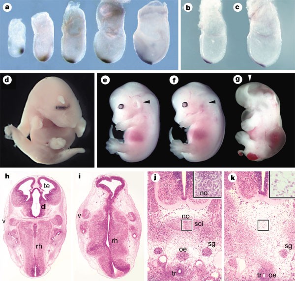
The organizer factors Chordin and Noggin are required for mouse forebrain development Nature volume 403, pages658–661 (2000) 10 February 2000 https://www.nature.com/articles/35001072
In vitro modelling of anterior primitive streak patterning with human pluripotent stem cells identifies the path to notochord progenitors Posted April 22, 2024. bioRxiv https://www.biorxiv.org/content/10.1101/2023.06.01.543323v3.full
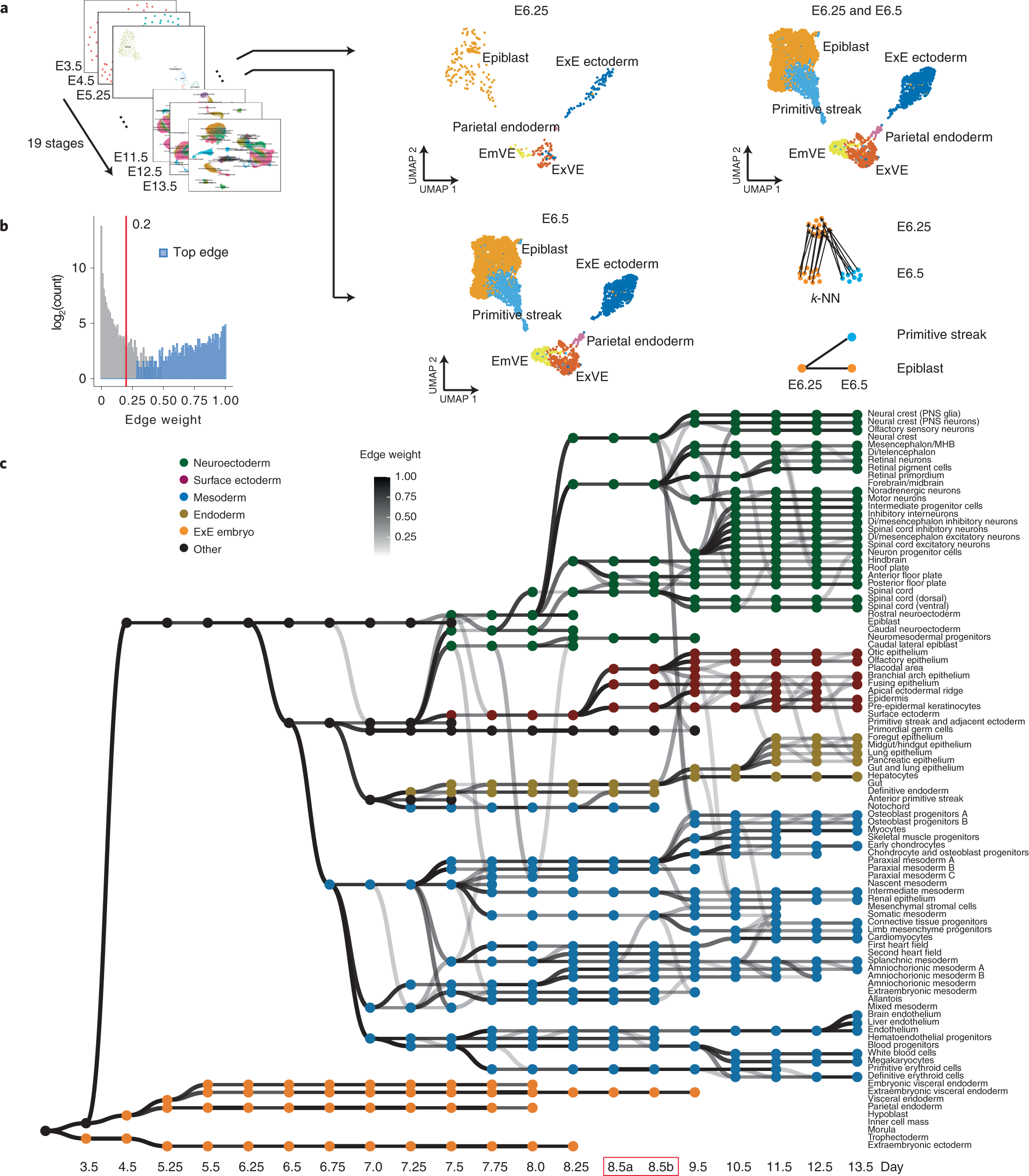
Recent advances in understanding cell types during human gastrulation Semin Cell Dev Biol. 2022 May 21;131:35–43. doi: 10.1016/j.semcdb.2022.05.004 https://pmc.ncbi.nlm.nih.gov/articles/PMC7615356/
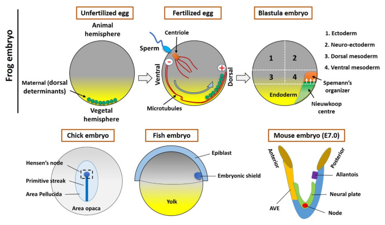
The Organizer and Its Signaling in Embryonic Development J Dev Biol. 2021 Nov 1;9(4):47. doi: 10.3390/jdb9040047 https://pmc.ncbi.nlm.nih.gov/articles/PMC8628936/ MDPI誌オープンアクセス論文
Mesoderm induction and patterning: insights from neuromesodermal progenitors Semin Cell Dev Biol. 2021 Nov 25;127:37–45. doi: 10.1016/j.semcdb.2021.11.010
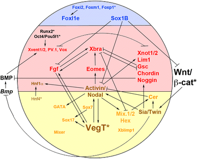
A gene regulatory program controlling early Xenopus mesendoderm formation: network conservation and motifs Semin Cell Dev Biol. 2017 Mar 22;66:12–24. doi: 10.1016/j.semcdb.2017.03.003
Vertebrate Axial Patterning: From Egg to Asymmetry Adv Exp Med Biol. 2017;953:209–306. doi: 10.1007/978-3-319-46095-6_6 https://pmc.ncbi.nlm.nih.gov/articles/PMC6550305/
Relationship between asymmetric nodal expression and the direction of embryonic turning Published: 09 May 1996 Jérôme Collignon, Isabella Varlet & Elizabeth J. Robertson Nature volume 381, pages155–158 (1996)
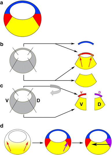
Molecular specification of germ layers in vertebrate embryos 14 December 2015 Cellular and Molecular Life Sciences Volume 73, pages 923–947, (2016) https://link.springer.com/article/10.1007/s00018-015-2092-y
The organization center of the amphibian embryo: its origin, spatial organization, and morphogenetic action P D Nieuwkoop PMID: 4581327 DOI: 10.1016/b978-0-12-028610-2.50005-8 Adv Morphog . 1973:10:1-39. doi: 10.1016/b978-0-12-028610-2.50005-8.
Fig. 1. (A) A secondary axis can be induced in developing Xenopus embryos by injection of RNA encoding β-catenin into a ventral cell of 4-cell stage embryos. Ventral cells are usually distinguished by their larger size and darker pigment compared to dorsal cells. For detailed methods see (Kuhl and Pandur, 2008a). (B) The duplicated axis is visible in neurula stage embryos within 2 days of injection. Embryos in these images have undergone in situ hybridisation for neuralβ-tubulin to illustrate the bilateral stripes of primary neurons and trigeminal https://www.researchgate.net/publication/272524241_An_oncologist%27s_friend_How_Xenopus_contributes_to_Cancer_research/figures
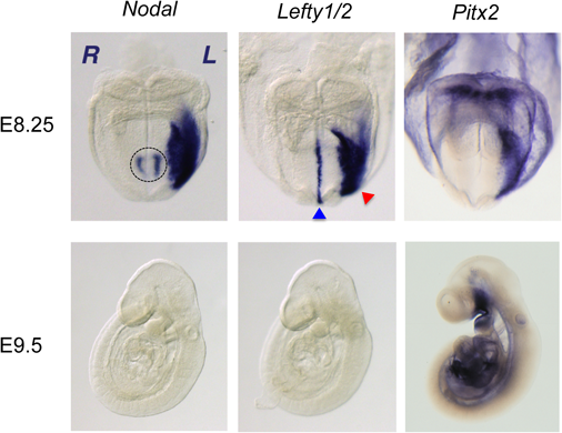
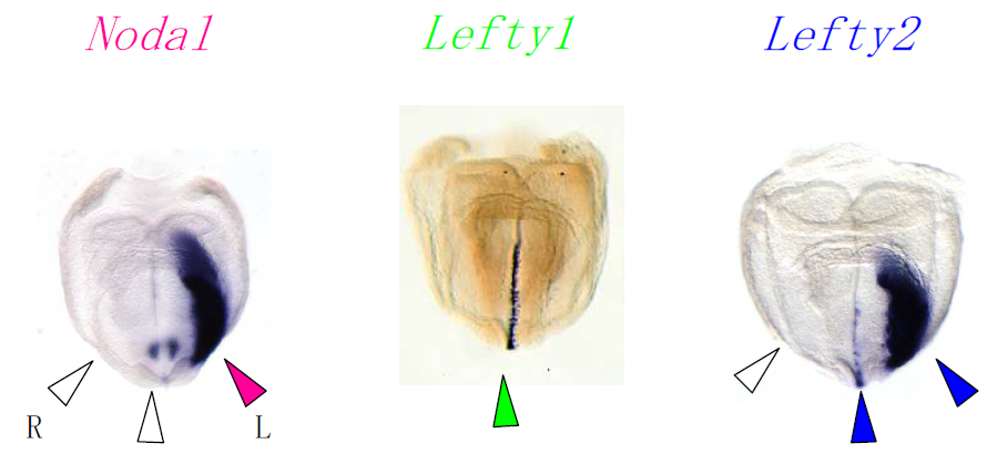
Left-right asymmetric expression of the TGF beta-family member lefty in mouse embryos C Meno 1, Y Saijoh, H Fujii, M Ikeda, T Yokoyama, M Yokoyama, Y Toyoda, H Hamada Nature . 1996May 9;381(6578):151-5. doi: 10.1038/381151a0. https://pubmed.ncbi.nlm.nih.gov/8610011/ 本文有料
総説
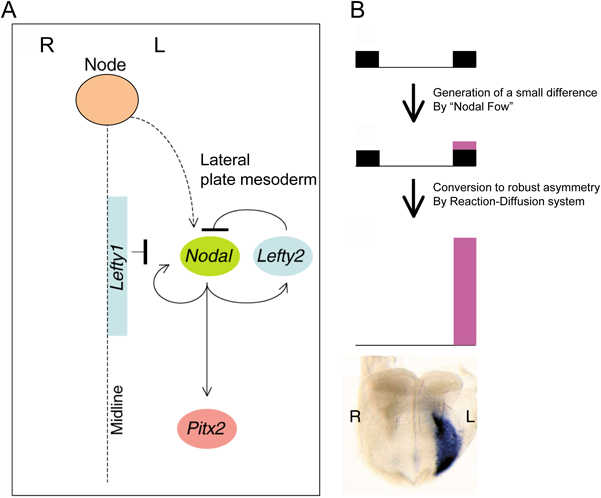
Molecular and cellular basis of left–right asymmetry in vertebrates Hiroshi HAMADA Proceedings of the Japan Acade …/Volume 96 (2020) Issue 7/Article overview/Full view Reviews PDF https://www.jstage.jst.go.jp/article/pjab/96/7/96_PJA9607B-04/_pdf/-char/en
Developmental Biology Volume 256, Issue 1, 1 April 2003, Pages 161-173 Developmental Biology Regular article Left–right patterning of the mouse lateral plate requires nodal produced in the node Author links open overlay panel Yukio Saijoh a , Shinya Oki a , Sachiko Ohishi a , Hiroshi Hamada a https://www.sciencedirect.com/science/article/pii/S0012160602001215
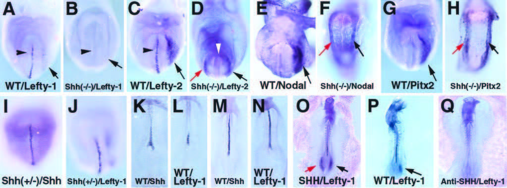
論文Multiple left-right asymmetry defects in Shh2/2 mutant mice unveil a convergence of the Shh and retinoic acid pathways in the control of Lefty-1 の図でもLefty-1は正中線上にしか局在していません。論文の記述(下)をよくよく読んだら、Lefty-1は将来floor plate(神経管の底板)になるところの左側に局在しているのだそうです。弱拡大の写真だとよくわかりませんでした。
FGF-induced vesicular release of Sonic hedgehog and retinoic acid in leftward nodal flow is critical for left–right determination Yosuke Tanaka, Yasushi Okada & Nobutaka Hirokawa Nature volume 435, pages172–177 (2005) Published: 12 May 2005
Multiple left-right asymmetry defects in Shh2/2 mutant mice unveil a convergence of the Shh and retinoic acid pathways in the control of Lefty-1 Proc. Natl. Acad. Sci. USAVol. 96, pp. 11376–11381, September 1999https://www.researchgate.net/publication/12800556_Multiple_Left-Right_Asymmetry_Defects_in_Shh–_Mutant_Mice_Unveil_a_Convergence_of_the_Shh_and_Retinoic_Acid_Pathways_in_the_Control_of_Lefty-1
An Additional Limb Can Be Induced from the Flank of the Chick Embryo by FGF4 Biochemical and Biophysical Research Communications Volume 209, Issue 3, 26 April 1995, Pages 809-816
肢芽 limb budの細胞は上皮間葉転換によって生じた間葉系細胞か
consensus.aiに訊いてみました。
The formation of vertebrate limb buds is a critical process in developmental biology, involving the transformation of specific cells to initiate limb development. This process is believed to be driven by epithelial-to-mesenchymal transition (EMT), where epithelial cells convert into mesenchymal cells, which are more migratory and capable of forming the limb structure.
Key Insights
Localized EMT Initiates Limb Bud Formation:
Limb buds form through a localized EMT of the coelomic epithelium within the presumptive limb fields, regulated by genes such as Tbx5 and Fgf1012.
The mesenchymal limb progenitors arise specifically from this EMT process, rather than from differential cell proliferation12.
Role of Lateral Plate Mesoderm (LPM):
The somatic LPM undergoes EMT to produce mesenchymal cells that contribute to limb bud formation. This process is driven by transcriptional regulators like PRRX1 and TWIST1, although further functional data are needed to fully understand these mechanisms2.
Epithelial-Mesenchymal Interactions:
Limb bud development involves interactions between the mesenchymal cells of the lateral plate mesoderm and the overlying ectodermal cells, highlighting the importance of epithelial-mesenchymal interactions in early morphogenesis45.
In Vitro Limb Bud Formation:
Limb bud-like tissues can be induced from murine pluripotent stem cells in vitro, demonstrating the potential to recreate the mesenchymal/epithelial complex tissues necessary for limb development3.
Conclusion
The origin of limb buds is indeed mesenchymal cells derived from epithelial-to-mesenchymal transition. This process is initiated by localized EMT in the coelomic epithelium and the somatic lateral plate mesoderm, regulated by specific genes and involving critical epithelial-mesenchymal interactions. These findings underscore the fundamental role of EMT in the early stages of limb bud formation.
Vertebrate Limb bud formation is initiated by localized Epithelial to Mesenchymal Transition Jerome Gros 1,#, Clifford J Tabin Science. 2014 Mar 14;343(6176):1253–1256. doi: 10.1126/science.1248228 https://pmc.ncbi.nlm.nih.gov/articles/PMC4097009/
Histone H3 lysine 4 methylation is mediated by Set1 and required for cell growth and rDNA silencing in Saccharomyces cerevisiae doi: 10.1101/gad.940201 Genes & Dev. 2001. 15: 3286-3295 https://genesdev.cshlp.org/content/15/24/3286.long
Strahl B.D., Ohba R., Cook R.G., Allis C.D.(1999) Methylation of histone H3 at lysine 4 is highly conserved and correlates with transcriptionally active nuclei in Tetrahymena. Proc. Natl. Acad. Sci. 96:14967–14972. https://www.pnas.org/doi/full/10.1073/pnas.96.26.14967
cDNAとは、mRNAを鋳型として逆転写酵素により合成したDNA(一重鎖)のことです。cDNAはcomplemantary DNA to mRNAという意味です。通常、実験するうえでcDNAを一重鎖のままにしておくことはなく、cDNAを鋳型としてDNA合成酵素を働かせて二重鎖を得ます。そうしないと、増やしたりできないので。最初につくった一本鎖のcDNAはアンチセンス鎖ですが、それを鋳型にして二重鎖にするために合成した方は、センス鎖になります。cDNAクローニングの論文で示される塩基配列は、センス鎖の塩基配列です。cDNAはmRNAを鋳型にして作った一本鎖DNAという言い方を見ると、論文でcDNA配列といったときの配列とは逆になるのでは?という疑問が湧いて、混乱を招きそうです。そのあたりを、ハッキリさせておきます。
ChatGPTに説明してもらいました。
cDNA is initially single-stranded after it is synthesized from mRNA, and there’s a subtle distinction when considering single- and double-stranded DNA.
Here’s a more precise breakdown of what happens:
Single-stranded cDNA:
cDNA is synthesized from the mRNA template by reverse transcriptase, making it complementary to the mRNA. Since mRNA is complementary to the anti-sense (template) strand of genomic DNA, the single-stranded cDNA is complementary to the mRNA, which essentially makes it analogous to the anti-sense strand of genomic DNA.
Double-stranded cDNA:
If the cDNA is made into a double-stranded molecule (which is often the case in practical applications), then the second strand is synthesized to be complementary to the first (single-stranded) cDNA. This newly synthesized strand is now complementary to the cDNA and, as a result, matches the sequence of the original mRNA (or the sense strand of genomic DNA).
Thus, when cDNA is single-stranded, it is effectively the anti-sense strand (complementary to mRNA). However, once you make it double-stranded, the complementary strand matches the sense strand of the original genomic DNA.
Summary:
Single-stranded cDNA corresponds to the anti-sense strand of the genomic DNA.
When cDNA becomes double-stranded, the newly synthesized second strand is the sense strand, which matches the original genomic DNA’s sense strand (with T instead of U).
The strand from the “first-strand synthesis” of cDNA is the one synthesized directly from the mRNA template. Since mRNA is complementary to the anti-sense strand of the original genomic DNA, this first-strand cDNA is complementary to the mRNA and is therefore analogous to the anti-sense strand of the original DNA.
The strand from the “second-strand synthesis” of cDNA is synthesized using the first-strand cDNA as a template. This second strand will be complementary to the first strand, which means it will have the same sequence as the original sense strand of the genomic DNA (except for thymine (T) replacing uracil (U) found in mRNA).
In summary:
The first-strand cDNA corresponds to the anti-sense strand.
The second-strand cDNA corresponds to the sense strand.
 (C)Expression of goosecoid in 8.0 dpc embryos of the indicated genotypes was analyzed by in situ hybridization. (D)Sections through the embryos in C.