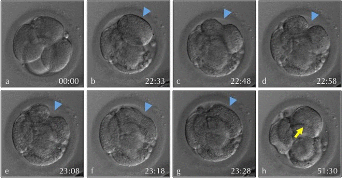
哺乳類の卵の発生において、卵割期にコンパクション compactionと言う現象が起きます。これは16細胞頃に外側の細胞が互いにぴったりと接着することにより、それまではまるっこい割球がくっついていただけの胚が、割球同士の境目がわからなくなるくらいに密着した状態に変化するものです。
- Analysis of compaction initiation in human embryos by using time-lapse cinematography Embryo Biology Open access Published: 09 March 2014 Volume 31, pages 421–426, (2014) https://link.springer.com/article/10.1007/s10815-014-0195-2

明らかに細胞接着因子がそこで活躍しているはずですし、外側の細胞と内側の細胞という2種類の細胞への分化という、一連の分化の流れの出発点にもなります。
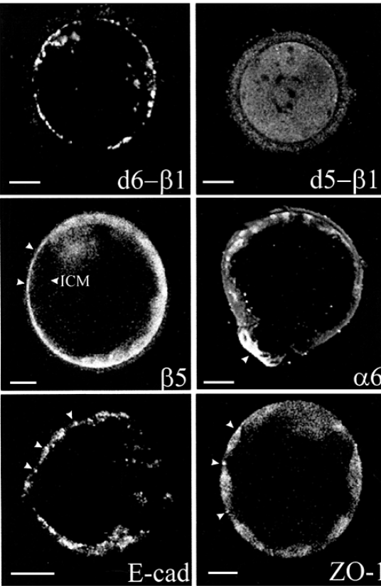
哺乳類の卵割の場合、最初の外見的な細胞分化は、コンパクションによって胚の表面部分に位置する細胞(互いが細胞接着をぴったりとしている)と、その内部に位置する細胞の2種類です。コンパクションのあと、外側の細胞はさらに強固に接着します。それは、タイトジャンクションが形成されることによります。
 https://www.sciencedirect.com/science/article/pii/S0005273607003562
Fig. 1. ZO-1 protein expression is stage-specific in developing mouse embryos freshly collected from the reproductive tracts during preimplantation period of pregnancy. https://www.sciencedirect.com/science/article/pii/S0012160608002121
human compaction
- Human Embryos Developing in Vitro are Susceptible to Impaired Epithelial Junction Biogenesis Correlating with Abnormal Metabolic Activity 2007 https://academic.oup.com/humrep/article/22/8/2214/645208

- Gene expression regulating epithelial intercellular junction biogenesis during human blastocyst development in vitro 2003年
- Expression of cell adhesion molecules during human preimplantation embryo development D J Bloor 1, A D Metcalfe, A Rutherford, D R Brison, S J Kimber Mol Hum Reprod . 2002 Mar;8(3):237-45. doi: 10.1093/molehr/8.3.237. 本文無料 PDF

- Compaction and surface polarity in the human embryo in vitro G Nikas 1, A Ao, R M Winston, A H Handyside Biol Reprod . 1996 Jul;55(1):32-7. doi: 10.1095/biolreprod55.1.32. 本文有料
タイトジャンクションとアドヘレンジャンクションとの関係性
上皮細胞においては,TJはアピカル膜直下の非常に限られた細胞膜の領域に形成されるが,TJの形成は,どのように制御されているのであろうか.上皮細胞の細胞接着を一度破壊し,その再形成過程を詳しく観察すると,TJの形成に先立って,まずE-カドヘリンを介したAJが形成される.この際に,E-カドヘリンの阻害抗体を細胞に処理すると,AJのみならずTJの形成も阻害される3).また,AJの必須の構成要素であるα-カテニンの発現が消失したPC-9細胞では,AJのみならずTJも形成されない4).このような観察事実から,TJ形成にはAJの形成が必要であることが明らかになった.その理由として,AJを形成することによって細胞膜どうしを物理的に近接させることによって,隣接細胞間のクローディンどうしの結合が可能となりTJの形成を可能にしているのではないかと考えられてきた.しかし近年になって,AJの形成は,細胞骨格の再組織化,低分子量Gタンパク質の活性化などのさまざまな変化を細胞内にもたらすことで,TJ形成に寄与することが示唆された.さらに,筆者らは最近,AJの形成が形質膜の脂質組成を変化させ,TJの形成を促進することを明らかにした5). https://seikagaku.jbsoc.or.jp/10.14952/SEIKAGAKU.2019.910555/data/index.html
参考
- Mechanical strengthening of cell-cell adhesion during mouse embryo compaction Ludmilla de Plater Julie Firmin Jean-Léon Maître Open AccessPublished:March 25, 2024DOI:https://doi.org/10.1016/j.bpj.2024.03.028 Compaction requires the cell-cell adhesion molecule Cdh1, also known as E-cadherin and initially called uvomorulin after it was discovered in compacting mouse embryos.
- Cell-cell adhesions in embryonic stem cells regulate the stability and transcriptional activity of β-catenin FEBS Lett. 2022 Jul; 596(13): 1647–1660. Published FEBS Lett. 2022 Jul; 596(13): 1647–1660. Published online 2022
- Cell-cell adhesions in embryonic stem cells regulate the stability and transcriptional activity of β-catenin Apr 6. doi: 10.1002/1873-3468.14341 PMCID: PMC10156795 NIHMSID: NIHMS1793009 PMID: 35344589
- Published: 06 August 2020 Transcriptomic analysis reveals differential gene expression, alternative splicing, and novel exons during mouse trophoblast stem cell differentiation Rahim Ullah, Ambreen Naz, Hafiza Sara Akram, Zakir Ullah, Muhammad Tariq, Aziz Mithani & Amir Faisal Stem Cell Research & Therapy volume 11, Article number: 342 (2020) https://stemcellres.biomedcentral.com/articles/10.1186/s13287-020-01848-8
- Volume 21, Issue 13, 26 December 2017, Pages 3957-3969 Journal home page for Cell Reports Resource Protein Expression Landscape of Mouse Embryos during Pre-implantation Development https://www.sciencedirect.com/science/article/pii/S2211124717317953
- How Adhesion Forms the Early Mammalian Embryo 2015White わかりやすい実験データの図およびまとめの細胞シグナルの図
- Making the first decision: lessons from the mouse Agnieszka Jedrusikcorresponding author 1 , 2 Reprod Med Biol. 2015 Oct; 14(4): 135–150. Published online 2015 Apr 16. doi: 10.1007/s12522-015-0206-8 PMCID: PMC5715835 PMID: 29259411 https://www.ncbi.nlm.nih.gov/pmc/articles/PMC5715835/
- RNA-Seq Transcriptome Profiling of Equine Inner Cell Mass and Trophectoderm1 Khursheed Iqbal, James L. Chitwood, Geraldine A. Meyers-Brown, Janet F. Roser, Pablo J. Ross Author Notes Biology of Reproduction, Volume 90, Issue 3, 1 March 2014, 61, 1-9, https://doi.org/10.1095/biolreprod.113.113928 Published: 01 March 2014
- Changes in sub-cellular localisation of trophoblast and inner cell mass specific transcription factors during bovine preimplantation development Zofia E Madeja, Jaroslaw Sosnowski, Kamila Hryniewicz, Ewelina Warzych, Piotr Pawlak, Natalia Rozwadowska, Berenika Plusa & Dorota Lechniak BMC Developmental Biology volume 13, Article number: 32 (2013) Published: 13 August 2013 https://bmcdevbiol.biomedcentral.com/articles/10.1186/1471-213X-13-32
- Cadherin-dependent filopodia control preimplantation embryo compaction Nature Cell Biology volume 15, pages1424–1433 (2013) Published: 24 November 2013
- 細胞の極性化から眺めたマウス着床前の形作りの仕組み 2012.02.28 基礎生物学研究所 https://www.nibb.ac.jp/pressroom/news/2012/02/28.html Prickle2タンパク質は、2細胞期から核で発現が開始し、8細胞期までは核内に局在。それ以降、胚盤胞期までは主に細胞質での粒子状の発現。 Prickle2遺伝子破壊マウスの表現型は、25細胞期以降に胚盤胞が形成されない、また、栄養外胚葉への分化マーカーの発現が劇的に減少。頂底軸に沿ったいくつかのマーカー分子の発現が異常。Prickle2遺伝子は8細胞期に起こる細胞の極性確立に重要。その後の細胞の運命決定、胚盤胞の形成にも関与。

- ORAL PRESENTATION | OTHER: ART| VOLUME 94, ISSUE 4, SUPPLEMENT , S21, SEPTEMBER 2010 PDF [42 KB] Save Share Reprints Request Differential gene expression profiles between human embryo on day-3 and trophoblast cells on day-5: specific molecular signature S. Assou D. Haouzi F. Pellestor H. Dechaud J. De Vos S. Hamamah DOI:https://doi.org/10.1016/j.fertnstert.2010.07.082 https://www.fertstert.org/article/S0015-0282(10)01187-8/fulltext
- Tight junction biogenesis during early development 2008年レビュー論文 https://www.sciencedirect.com/science/article/pii/S0005273607003562
- Development . 2004 Dec;131(23):5817-24. doi: 10.1242/dev.01458. Epub 2004 Nov 3. Stabilization of beta-catenin in the mouse zygote leads to premature epithelial-mesenchymal transition in the epiblast https://pubmed.ncbi.nlm.nih.gov/15525667/
- 15 SEPTEMBER 2004 Maternal β-catenin and E-cadherin in mouse development Wilhelmine N. de Vries, Alexei V. Evsikov, Bryce E. Haac, Karen S. Fancher, Andrea E. Holbrook, Rolf Kemler, Davor Solter, Barbara B. Knowles Author and article information Development (2004) 131 (18): 4435–4445. https://journals.biologists.com/dev/article/131/18/4435/42290/Maternal-catenin-and-E-cadherin-in-mouse
- Regulation of cell adhesion during embryonic compaction of mammalian embryos: roles for PKC and beta-catenin C M Pauken 1, D G Capco Mol Reprod Dev . 1999 Oct;54(2):135-44. doi: 10.1002/(SICI)1098-2795(199910)54:2<135::AID-MRD5>3.0.CO;2-A. https://pubmed.ncbi.nlm.nih.gov/10471473/
参考
aPKC
- Inactivation of aPKCλ Reveals a Context Dependent Allocation of Cell Lineages in Preimplantation Mouse Embryos Nicolas Dard ,Tran Le,Bernard Maro,Sophie Louvet-Vallée Published: September 21, 2009 https://doi.org/10.1371/journal.pone.0007117 https://journals.plos.org/plosone/article?id=10.1371/journal.pone.0007117
カテニンの細胞接着における役割
- Cadherins:single transmembrane domain glycoproteins that mediate calcium-dependent cell–cell adhesion via homophilic interactions
- The intracellular domain of E-cadherin associates with a protein family collectively termed catenins.
- β-catenin and γ-catenin (plakoglobin) interact directly with E-cadherin’s COOH-terminal domain in a mutually exclusive way, and both proteins associate with α-catenin, which links the cadherin complexes to the actin cytoskeleton and mediates stable cell adhesion.
- p120ctn is another catenin family member, which binds to the cytoplasmic juxtamembrane portion of E-cadherin and influences E-cadherin clustering and adhesive strength.
カテニンのwntシグナリングにおける役割
- wnt不在のとき:細胞質β-cateninは axin/conductinやadenomatous polyposis coli tumor suppressor proteinと複合体をつくり、 glycogen synthase kinase 3βによってN末がリン酸化を受けておりプロテオソームによる分解を受ける。
- wntが受容体frizzledに結合すると、一連のシグナル経路によってglycogen synthase kinase 3βによるβ-cateninリン酸化が抑制され、β-cateninは安定化。
- 安定化したβ-cateninは、high mobility group (HMG) box転写因子であるlymphoid enhancer binding factor (LEF)-1/T cell factor (TCF) ファミリーと結合する.
- 核内β-catenin–LEF/TCF複合体はhistone acetylase p300/CBPなどのコアクチベーターと結合して、TATA-binding protein TBPの発現を促進したり, groucho-related gene 5 などとの結合により遺伝子発現を抑制したりする。
(とある論文のイントロ部分のまとめ)
- OCT4 Coordinates with WNT Signaling to Pre-pattern Chromatin at the SOX17 Locus during Human ES Cell Differentiation into Definitive Endoderm Stem Cell Reports. 2015 Oct 13; 5(4): 490–498. Published online 2015 Sep 24. doi: 10.1016/j.stemcr.2015.08.014 PMCID: PMC4624996 PMID: 26411902
- The E-Cadherin and N-Cadherin Switch in Epithelial-to-Mesenchymal Transition: Signaling, Therapeutic Implications, and Challenges Cells. 2019 Oct; 8(10): 1118. Published online 2019 Sep 20. doi: 10.3390/cells8101118 PMCID: PMC6830116 PMID: 31547193 https://www.ncbi.nlm.nih.gov/pmc/articles/PMC6830116/

その他の参考
- https://www.sciencedirect.com/science/article/pii/S0925477300004160
- https://www.sciencedirect.com/science/article/pii/S0012160607013991
