体細胞分裂における染色体分離の概観
Textbook picture of the spindle. Redrawn and modified from (Alberts et al. 2014)
https://www.ncbi.nlm.nih.gov/pmc/articles/PMC5845649/


中心体
Kinetochore fibers. Electron micrograph of a metaphase spindle in a PtK1 cell. Kinetochore microtubules are visible as thin lines extending between the boundary of the spindle pole (curved dashed line) and the kinetochores (K1–K5). Arrows mark microtubules that leave the plane of section; V vesicles, PCM pericentriolar material; scale bar 0.5 µm. Image reproduced with permission from (McDonald et al. 1992)
https://www.ncbi.nlm.nih.gov/pmc/articles/PMC5845649/


中心小体centrioleは、小さな筒みたいな装置で、3本の微小管がトリプレットになって、そのトリプレットが9個並んで、筒のような形をしています。
中心小体が二個一組、相互に直角対向しL字形に配置している構造が、中心体(ちゅうしんたい、centrosome)で、微小管形成中心(MTOC; microtubule organizing center)とも呼ばれます。なお植物細胞においては中心体は存在しません。それでも植物細胞は細胞分裂をすることができるので不思議です。(植物細胞の)細胞分裂では、中心小体が必須というわけではないのでしょうか。
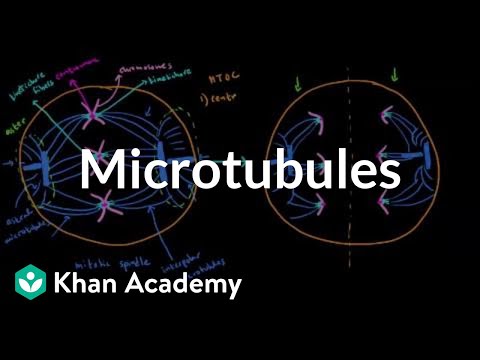
Microtubules | Cells | MCAT | Khan Academy khanacademymedicine チャンネル登録者数 179万人
紡錘体
Spindle microtubules can be divided into three major classes:
- kinetochore microtubules, which form k-fibers ending at the kinetochore;
- interpolar microtubules, which extend from the opposite sides of the spindle and interact in the middle; and
- astral microtubules, which extend towards the cell cortex.
https://link.springer.com/article/10.1007/s00249-017-1244-4

Interpolar microtubules
https://www.nature.com/scitable/content/types-of-microtubules-involved-in-mitosis-14752887/


中心体と中心体とを結ぶ微小管が、染色体分離のための力を生み出しているのかなと思ったのですが、そう単純ではないようです。下の説明だとむしろ安定的に保持しているような仮説になっていました。かりにそうだとしても、染色体を中心体の方に集結させる必要があるので、やはりmitotic spindle(染色体に結合した紡錘糸)が染色体を引き寄せる必要があります。
MAPs (microtubule associated proteins) crosslink antiparallel interpolar microtubules to create a stable midzone that allows kinesin motor proteins to generate sliding forces that push the spindle poles apart (4).
The importance of microtubule-dependent tension in accurate chromosome segregation Angela R. Bunning and Mohan L. Gupta Jr.corresponding author* Front Cell Dev Biol. 2023; 11: 1096333. Published online 2023 Jan 23. doi: 10.3389/fcell.2023.1096333 PMCID: PMC9899852 PMID: 36755973
https://www.ncbi.nlm.nih.gov/pmc/articles/PMC9899852/


- Interpolar microtubules are dispensable in fission yeast meiosis II Takashi Akera, Masamitsu Sato & Masayuki Yamamoto Nature Communications volume 3, Article number: 695 (2012) Published: 28 February 2012 https://www.nature.com/articles/ncomms1725 The mitotic spindle consists of two types of microtubules. Dynamic kinetochore microtubules capture kinetochores, whereas stable interpolar microtubules serve as the structural backbone that connects the two spindle poles. Both have been believed to be indispensable for cell division in eukaryotes.
J Cell Biol. 2017 Jun 5; 216(6): 1525–1531. doi: 10.1083/jcb.201612064 PMCID: PMC5461028 PMID: 28490474 Review The mechanics of microtubule networks in cell division Scott Forth and Tarun M. Kapoorcorresponding author https://www.ncbi.nlm.nih.gov/pmc/articles/PMC5461028/


- Mechanical Mechanisms of Chromosome Segregation Cells 2021, 10, 465. https://doi.org/10.3390/cells10020465 https://www.ncbi.nlm.nih.gov/pmc/articles/PMC7926803/pdf/cells-10-00465.pdf
- Motor function in interpolar microtubules during metaphase Author links open overlay panel J.M. Deutsch , Ian P. Lewis Journal of Theoretical Biology Volume 370 , 7 April 2015, Pages 1-10 Journal of Theoretical Biology https://www.sciencedirect.com/science/article/abs/pii/S002251931500020X
キネトコア 動原体 の構造と構成要素
- 定説を覆す!染色体の分配のしくみに、鍵となる新たな分子の働きを発見 新しい抗癌剤開発に期待 2018-11-13 大阪大学 大阪大学大学院生命機能研究科の深川竜郎教授・原昌稔助教らの研究グループは、染色体とその分裂装置である紡錘体との結合に関して、これまでの定説を覆してCENP-Tというタンパク質が関わっていることを世界で初めて明らかにしました。 染色体 とその分裂装置である紡錘体 との結合に関して、これまで、紡錘体と結合するための染色体上の構造であるキネトコア(動原体) では、CENP-C と呼ばれるタンパク質が重要と考えられていた。 ・CENP-T の制御メカニズムの詳細な解析により、CENP-CよりもCENP-Tが染色体の分配時に主要な役割を担っている
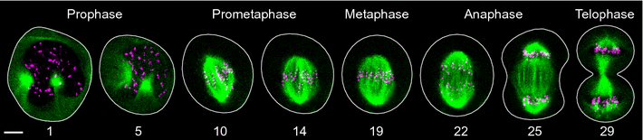
分裂後期(anaphase)における染色体分離のチェックポイント
Double-checking chromosome segregation Helder Maiato ORCID logo , Sónia Silva ORCID J Cell Biol (2023) 222 (5): e202301106. April 05 2023 https://doi.org/10.1083/jcb.202301106
https://rupress.org/jcb/article/222/5/e202301106/214000/Double-checking-chromosome-segregationDouble

