眼の発生の講義動画
- 3D Development of the Eye: A Comprehensive Overview – Eye Embryology – Ophthalmology MedicoVisual – Visual Medical Lectures チャンネル登録者数 4.26万人
眼の発生は、予定眼領域 eye fieldが生じるところから始まります。簡単に概略をいうと、
- 予定眼領域 eye fieldが形成される
- 予定眼領域が左右に分かれる(突然変異体などで、分かれなかった場合は、一つ眼になってしまう!)
- 眼胞(がんぽう)が、形成される。このころはまだ前脳の部分の神経管は閉じていません。ですから、最初は、神経溝の一部から隆起して眼胞が形成されることになります。
The cellular and molecular mechanisms of vertebrate lens development November 2014 Development 141(23):4432-4447 DOI: 10.1242/dev.107953 CC BY 3.0 https://www.researchgate.net/publication/268985222_The_cellular_and_molecular_mechanisms_of_vertebrate_lens_development#fullTextFileContent
眼の発生の分子メカニズム
眼のマスター遺伝子による予定眼領域 eye filedの確立
脳の前方ではWnt/β-カテニン シグナル経路は抑制されており(Dkkなどにより)、そのかわりノンカノニカルなWntシグナルがeye filedを確立するのに重要な働きをするようです。下の図のようにWNTがeye filedになるすぐ後ろ側から分泌されて、ノンカノニカル経路が働くことで眼のマスター遺伝子であるRAXやPax6の遺伝子発現が誘導されます。
Wnt signaling in eye organogenesis Sabine Fuhrmann Organogenesis Pages 60-67 | Published online: 11 Jun 2008 Cite this article https://doi.org/10.4161/org.4.2.5850 https://www.tandfonline.com/doi/full/10.4161/org.4.2.5850#d1e615
RAX
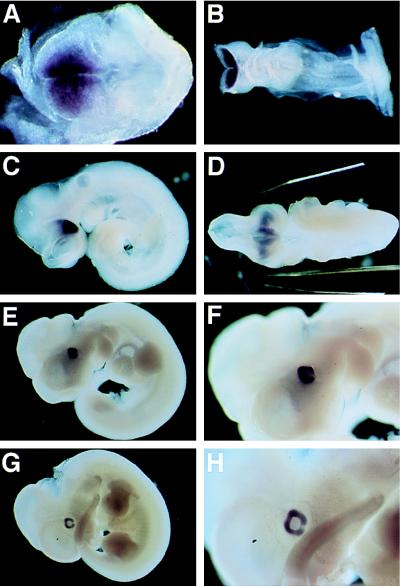
眼ののマスター遺伝子といえばPAX6が有名です。PAX6をショウジョウバエで強制発現させると発現させた場所に眼が生じることが示されています。しかしマウスにおいてはPAX6遺伝子の働きが無くても眼が形成されるそうです。RAXという転写因子がその後見つかっており、こちらのほうがPAXよりも早くから発現することが知られています。
Whole-mount in situ hybridization of E7.5 (A), E8.5 (B), E9.5 (C and D), E10.5 (E and F), and E11.5 mouse embryos (G and H). rax, a novel paired-type homeobox gene, shows expression in the anterior neural fold and developing retina Takahisa Furukawa *, Christine A Kozak †, Constance L Cepko *,‡ Proc Natl Acad Sci U S A. 1997 Apr 1;94(7):3088–3093. doi: 10.1073/pnas.94.7.3088 ”pax6 expression starts later than that of rax, suggesting that rax might be directly or indirectly upstream of pax6 in the series of events that lead to optic vesicle formation.” https://pmc.ncbi.nlm.nih.gov/articles/PMC20326/
RAX遺伝子破壊マウスでは眼の形成ができません。
An essential role for Rax in retina and neuroendocrine system development Yuki Muranishi, Koji Terada, Takahisa Furukawa First published: 24 April 2012 https://doi.org/10.1111/j.1440-169X.2012.01337.x DGD https://onlinelibrary.wiley.com/doi/10.1111/j.1440-169X.2012.01337.x
PAX6
眼の発生のマスター遺伝子の一つであるPAX6を欠損させたマウスでは眼が全くできなくなります。
Anophthalmia mouse mutant. a Head of a neonatal (P1) homozygous Pax6Aey11 mutant compared to a wild-type mouse (wt) at the same age. The absence of eyes in the mutant is obvious. The eyelids of neonatal mice are still closed (photography: Jana Löster†, unpublished). Mouse models for microphthalmia, anophthalmia and cataracts 27 March 2019 Volume 138, pages 1007–1018, (2019)https://link.springer.com/article/10.1007/s00439-019-01995-w
カエルの眼の発生に関わるマスター遺伝子の発現パターン
Specification of the vertebrate eye by a network of eye field transcription factors Michael E. Zuber, Gaia Gestri, Andrea S. Viczian, Giuseppina Barsacchi, William A. Harris Author and article information Development (2003) 130 (21): 5155–5167. https://journals.biologists.com/dev/article/130/21/5155/52150/Specification-of-the-vertebrate-eye-by-a-network
予定眼領域 eye filedの左右への分離
正中線シグナルであるshhが正中線で分泌される結果PAX2の発現が誘導されます。PAX2はPAX6を抑制することにより、真ん中部分は予定眼領域ではなくなり、予定眼領域が左右に分かれます。
眼胞によるレンズプラコードの誘導の誘導
眼胞からBMPやFGFなどのシグナル分子が分泌されて外胚葉に働きかけ、レンズを誘導します。
Figure 2. Expression of Bmp4 and BMP type-I receptor genes during early eye development. (A–F) In situ hybridization using an antisense riboprobe for Bmp4 on transverse sections of 10- (A) and 14- (B) somite-stage embryos, and on frontal sections of 18- (C), 22- (D), 27- (E), and ∼40-somite-stage (10.5 dpc) (F) embryos. BMP4 is essential for lens induction in the mouse embryo Yasuhide Furuta and Brigid L.M. Hogan1 Genes & Dev. 1998. 12: 3764-3775 https://genesdev.cshlp.org/content/12/23/3764.full
Figure 3. The timing and intensity of FGF signalling controls the two-dimensional patterning of the lens. The PPR is first selected from the head ectoderm by active FGF signalling devoid of suppressive BMP and Wnt (a), before progressing further towards the LP fate in lieu of(~の代わりに◆instead of に近い意味) continuous FGF signalling (b). FGF next induces Frs2–Shp2-mediated Ras signalling modulated by NF1 to promote Pax6 expression and lens vesicle invagination (c), but FGF signalling must be suppressed by Spry to allow lens vesicle closure (d). During the subsequent lens maturation, FGF cooperates with PDGF to stimulate Notch signalling, which promotes lens epithelium proliferation (e). In lens fibre cells, FGF signalling also activates Ras to promote differentiation and recruit Ras and Rac GTPases via Crk/CrkL to promote cell elongation (f).
レンズの前後軸の決定に関わる分泌シグナル:WNTとFGF
Fig. 2. Diagram indicating how the ocular media and a gradient of FGF stimulation may determine antero-posterior patterns of lens cell behavior. Growth factor regulation of lens development F.J. Lovicu , J.W. McAvoy Developmental Biology Volume 280, Issue 1, 1 April 2005, Pages 1-14 https://www.sciencedirect.com/science/article/pii/S001216060500045X?via%3Dihub
眼胞から眼杯へ
眼胞から眼杯に形態が変わるときに重要な遺伝子として転写因子Lhx2が同定されています。下の論文によれば、Lhx2ノックアウトマウスでは眼胞までは形成されますが眼杯はできないそうです。また外胚葉がレンズに誘導される現象も起きないのだそうです。下の図が示すようにLhx2がBMPを亢進して、BMPがレンズ誘導や眼杯形成を促進するという仮説が提唱されています。
Lhx2 links the intrinsic and extrinsic factors that control optic cup formation Sanghee Yun, Yukio Saijoh, Karla E. Hirokawa, Daniel Kopinke, L. Charles Murtaugh, Edwin S. Monuki, Edward M. Levine Author and article information Development (2009) 136 (23): 3895–3906. https://journals.biologists.com/dev/article/136/23/3895/43745/Lhx2-links-the-intrinsic-and-extrinsic-factors
眼の発生の分子メカニズムのまとめ
下のレビュー論文の図が、眼の発生に関与するシグナル分子や転写因子を網羅的にまとまっていてわかりやすいと思います。
FIGURE 4 | Early ocular morphogenesis. The Use of Induced Pluripotent Stem Cells as a Model for Developmental Eye Disorders July 2020 Frontiers in Cellular Neuroscience 14:265 DOI: 10.3389/fncel.2020.00265 License CC BY (A) Developmental pathways such as Wnt, BMP, and fibroblast growth factor (FGF) drive upregulation of eye-field transcription factors in the anterior neural plate, creating the specified region known as the “eye-field.”
症例報告
- Cyclopia, a newborn with a single eye, a rare but lethal congenital anomaly: A case report Int J Surg Case Rep. 2021 Nov; 88: 106548. Published online 2021 Nov 4. doi: 10.1016/j.ijscr.2021.106548 PMCID: PMC8581486 PMID: 34741865
