心電図の詳細
ECG Simplified #1 Cardiac dipole and Instantaneous vector The Physiology Channel チャンネル登録者数 1.23万
ECG and Electric Dipoles UCL Medical Physics and Biomedical Engineering チャンネル登録者数 3440人
- https://www.bem.fi/ 12-Lead ECG System https://www.bem.fi/book/15/15.htm Bioelectromagnetism(本のPDF)https://www.bem.fi/book/book.pdf
- Physiological basis of electrocardiography By Alex Yartsev – 20/01/2018 Last updated 25/11/2024 – 14:43 https://derangedphysiology.com/main/cicm-primary-exam/cardiovascular-system/Chapter-735/physiological-basis-electrocardiography
- The ECG Curve: What Is It and How Does It Originate? https://thoracickey.com/2-the-ecg-curve-what-is-it-and-how-does-it-originate/
AP End − AP Epi = ECG
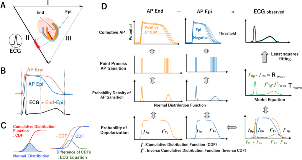 Fig 1. A. Asymmetry of ventricular wall and AP propagating from endocardial side to epicardial side and repolarization from epi to end generating R and T waves of ECG (lead II Positive Waves). B. Relationship between the cardiac dipole model and ECG: Depolarization starts from the endocardial side of ventricle muscle (Epi) and spreads to the epicardial side (End). Since the duration of the action potential is shorter on the Epi than the End., repolarization begins on the Epi and terminates on the End. The difference between the total potential on the Epi and that on the End of the myocardium is approximately equal to the ECG waveform. D. End and Epi AP elicit positive and negative potential, respectively. Their difference is closely related to ECG.
- Tensor cardiography: A novel ECG analysis of deviations in collective myocardial action potential transitions based on point processes and cumulative distribution functions Shingo Tsukada ,Yu-ki Iwasaki,Yayoi Tetsuo Tsukada Published: August 8, 2024 https://doi.org/10.1371/journal.pdig.0000273 https://journals.plos.org/digitalhealth/article?id=10.1371/journal.pdig.0000273
# 左室は内膜(endocardium)から外膜(epicardium)へ脱分極が進み、再分極は逆向きに起こるため、内層は正極性、外層は負極性の電位を ECG に与えます。結果として ECG 波形は「内層 AP − 外層 AP」の差分でほぼ説明できる.したがって「AP End − AP Epi = ECG」
左室心筋の**脱分極**は「内膜(endocardium)から外膜(epicardium)へ」進行し、**再分極**はその逆、「外膜から内膜へ」進みます[^2][^7][^9]。このため、**内膜側の活動電位(AP Endo)は外膜側(AP Epi)よりも持続が長く、再分極のタイミングが遅い**という特徴があります[^2][^9]。
心電図(ECG)は、**心筋壁内の電位差**、つまり「内膜側の電位−外膜側の電位」の変化を体表面で記録したものです[^4][^7]。この原理から、**ECG波形はおおむね「AP Endo − AP Epi」で説明できる**、すなわち
> **ECG ≈ AP Endo − AP Epi**
という関係が成立します[^4][^7]。
– **脱分極時**は内膜側が先に+(陽性)になり、その後外膜側が+になるため、電位差が生じてQRS波が形成されます[^7]。
– **再分極時**は外膜側が先に−(陰性)になり、内膜側が遅れて−になるため、T波が形成されます[^2][^7][^9]。
このように、**心電図の波形は「内膜AP − 外膜AP」の差分でほぼ説明できる**という理解は、現代の電気生理学的知見と合致しています[^4][^7]。
> 「心電図は、心内膜側の電位−心外膜側の電位=心電図ということは覚えておきましょう」[^7]
この考え方は、特に左室のような壁厚がある心筋で顕著です。
<div style=”text-align: center”>⁂</div>
[^1]: https://new.jhrs.or.jp/pdf/education/akiyamalecture09.pdf
[^2]: https://www.jstage.jst.go.jp/article/jse/43/3/43_201/_pdf/-char/ja
[^3]: https://new.jhrs.or.jp/pdf/education/akiyamalecture05.pdf
[^4]: https://www.jstage.jst.go.jp/article/jjsca/35/4/35_447/_pdf
[^5]: https://kenkyuukai.m3.com/journal/FilePreview_Journal.asp?path=sys%5Cjournal%5C20120523135602-83CF7C8E825B618D9952752533B880984CF5BE735C167A87E66E28B1AB28BC27.pdf\&sid=507\&id=202\&sub_id=2569
[^6]: https://www.jstage.jst.go.jp/article/jsmbe1963/23/7/23_7_452/_pdf
[^7]: https://www.byomie.com/wp-content/digitalBook/sampleVol2/pageindices/index7.html
[^8]: https://www.kango-roo.com/learning/2229/
[^9]: https://www.jamt.or.jp/studysession/center/asset/docs/基礎電気生理から心電図を学ぶ151010.pdf
[^10]: https://www.jstage.jst.go.jp/article/jsvc/47/1/47_1/_pdf
(https://www.perplexity.ai/)