臍からおしっこが漏れ出すことがあるというのを教科書で読んで、臍と膀胱がつながっているのかと感銘を受けたのですが、あれ?卵黄嚢も臍帯に含まれていなかったっけ?と疑問が生じました。もしそうなら腸管と臍が繋がるんじゃないかという疑問が湧いたわけです。
調べてみたら、そういうことはありませんでした。
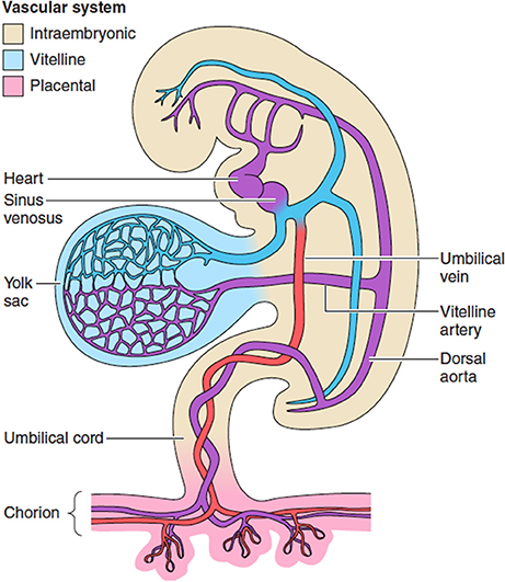
発生初期の胚の図を見ると卵黄嚢も臍帯に含まれているような描き方がされていると思います。しかしそれは早期だけのようです。”primitive umbilical cord”と、”definitive umbilical cord”というように言葉も使い分けられていました。”primitive umbilical cord”は umbilical ringとも言われるようです。
primitive umbilical cord
下のラングマンの教科書からの図では、腸管の伸長に伴う生理的なヘルニアが生じている時期を示しているので、断面図で腸管が描かれています(これは一時的なもの)。
Primitive umbilical cord and contents. A. A 5-week embryo showing structures passing through the primitive umbilical ring. B. The primitive umbilical cord of a 10-week embryo. C. Transverse section through the structures at the level of the umbilical ring. D. Transverse section through the primitive umbilical cord showing intestinal loops protruding in the cord. (Reprinted with permission from Sadler TW. Langman’s Medical Embryology. 13th ed. Philadelphia, PA: Wolters Kluwer Health; 2015.) https://renaissance.stonybrookmedicine.edu/system/files/Umbilical%20Cord%20Disorders.pdf
definitive umbilical cord もしくは単にumbilical cord 臍帯
Diagrams showing the (A) folding of the embryo and formation of the primitive umbilical cord and (B) definitive umbilical cord.
https://www.researchgate.net/publication/305720901_Anatomy_and_embryology_of_umbilicus_in_newborns_a_review_and_clinical_correlations
上の図では発生の早期と後期を示していて、一目瞭然でわかりやすいと思います。
https://www.frontiersin.org/journals/physiology/articles/10.3389/fphys.2018.00373/full
https://www.researchgate.net/publication/363584025_Bovine_omphalocele_errors_in_embryonic_development_veterinarian_importance_and_the_way_forward
https://www.researchgate.net/publication/301814976_Abdominal_Wall_and_Umbilicus-_Essentials_of_surgery-_Sunil_Chumber
Allantoi 尿膜の発生のその後
以下、ChatGPT4oによるまとめです。
The allantois is functionally obliterated early in development but leaves remnants that persist after birth. Here’s how it works:
Timeline of Allantois Obliteration 消滅:
- Early Embryogenesis:
- The allantois emerges as a small endodermal outpouching of the hindgut.
- It extends into the connecting stalk (which contributes to the formation of the umbilical cord) and plays a role in early gas exchange and the development of the umbilical arteries and veins.
- By the end of the first trimester, its role in development diminishes as the placenta takes over waste exchange.
- Obliteration during Development:
- The lumen of the allantois is obliterated during the second trimester, and it becomes a fibrous cord known as the urachus(尿膜管), which runs from the bladder to the umbilicus.
- By birth, the allantois is no longer a functional structure, but its remnant persists as the median umbilical ligament (正中臍索)in postnatal life.
- Postnatal Remnants:
- The median umbilical ligament extends from the apex of the bladder to the umbilicus and is the fibrous remnant of the allantois.
- If the obliteration of the allantois is incomplete, it can result in abnormalities like a urachal cyst, urachal sinus, or even a urachal fistula, which might require surgical correction.


