頭蓋骨の解剖学
下の動画は、非常にすっきりと説明してくれます。3Dイラストがカラフルで見やすく、頭に入りやすいです。
BONES OF THE SKULL – LEARN IN 4 MINUTES Neural Academy チャンネル登録者数 65.3万人
四肢の形成
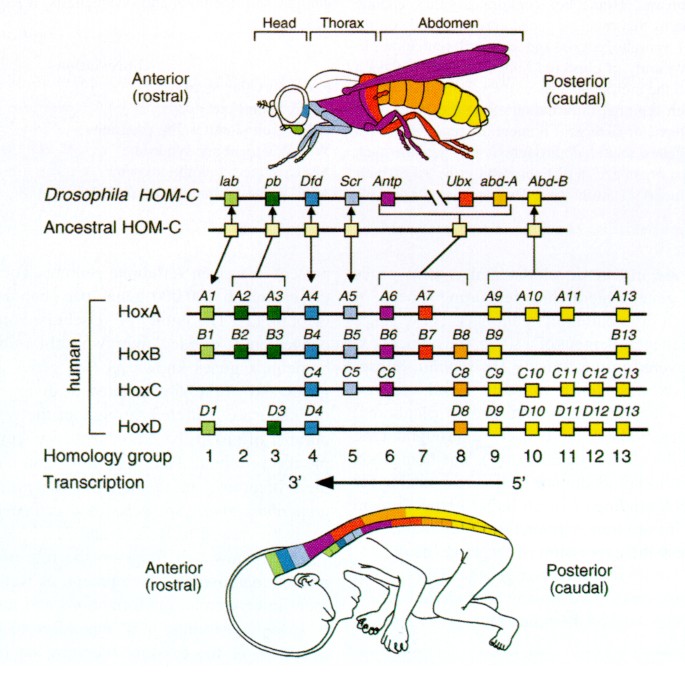
体軸方向のHOX遺伝子の発現
- Mark, M., Rijli, F. & Chambon, P. Homeobox Genes in Embryogenesis and Pathogenesis. Pediatr Res 42, 421–429 (1997). https://doi.org/10.1203/00006450-199710000-00001
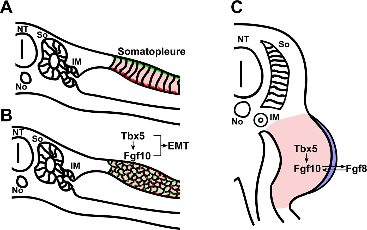
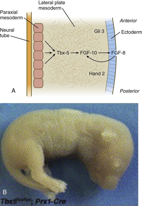
四肢の位置決めに関わる細胞シグナル
- Limb positioning and initiation: an evolutionary context of pattern and formation Dev Dyn. Author manuscript; available in PMC 2023 Nov 3. Published in final edited form as: Dev Dyn. 2021 Sep; 250(9): 1264–1279. Published online 2021 Feb 15. doi: 10.1002/dvdy.308 PMCID: PMC10623539
- Timed Collinear Activation of Hox Genes during Gastrulation Controls the Avian Forelimb Position Chloe Moreau Paolo Caldarelli Didier Rocancourt Nicolas Denans Olivier Pourquie Jerome Gros December 13, 2018 DOI:https://doi.org/10.1016/j.cub.2018.11.009 Current Biology VOLUME 29, ISSUE 1, P35-50.E4, JANUARY 07, 2019
- Review Article Molecular and evolutionary basis of limb field specification and limb initiation Mikiko Tanaka Develop. Growth Differ. (2013) 55, 149–163 (PDF) In tetrapods, motoneurons that innervate the limbs form lateral motor columns (LMCs) at the brachial and lumbar levels of the spinal cord, and the LMC identities in opposite forelimbs and hindlimbs are defined by expression of Hox6 and Hox10, respectively, in the spinal cord (Dasen et al. 2003; Shah et al. 2004; Wu et al. 2008).
- Hox9 genes and vertebrate limb specification. Cohn, M., Patel, K., Krumlauf, R. et al. Nature 387, 97–101 (1997). https://doi.org/10.1038/387097a0 無料要旨 Hox genes are good candidates for encoding position in lateral plate mesoderm along the body axis and thus for determining where limbs are formed. Local application of fibroblast growth factors (FGFs) to the anterior prospective flank of a chick embryo induces development of an ectopic wing, and FGF applied to posterior flank induces an ectopic leg.
肢芽の伸長に関わる分子シグナル
-
CHAPTER 10 Limb Development 13/06/2015
-
The mesenchymal factor, FGF10, initiates and maintains the outgrowth of the chick limb bud through interaction with FGF8, an apical ectodermal factor Hideyo Ohuchi, Takashi Nakagawa, Atsuyo Yamamoto, Akihiro Araga, Takeshi O 01 JUNE 1997 DEVELOPMENT チックのデータとモデル図
- Fibroblast Growth Factor 10 and Vertebrate Limb Development Libo Jin,1 Jin Wu,1 Saverio Bellusci,1,2,3,* and Jin-San Zhang1,2,4,* Front Genet. 2018; 9: 705. Published online 2019 Jan 7. doi: 10.3389/fgene.2018.00705 PMCID: PMC6338048 PMID: 30687387

- Initiation of Vertebrate Limb Deveiopment (PDF) Martin J. Cohn Thesis submitted for the degree of Ph. D
- J:71694 Bruneau S, et al., Dev Biol. 2001 Sep 15;237(2):345-53 https://www.informatics.jax.org/image/MGI:2153107

FGF8とFGF4の関係
こういう論文があります。
Biochemical and Biophysical Research Communications Volume 209, Issue 3, 26 April 1995, Pages 809-816 Biochemical and Biophysical Research Communications Regular Article An Additional Limb Can Be Induced from the Flank of the Chick Embryo by FGF4
ここではFGF4産生細胞が、異所性の肢芽の形成を引き起こすことを示す実験に使われています。しかし、内在性のFGF4は肢芽ではあまり発現していないようです。(AER).AERに発現するのはFGF8で、FGF8のノックアウトではFGF4が代償的に発現するようです。
BMP signals control limb interdigital programmed cell death by regulating FGF signaling Save Related Papers Chat with paper July 2007Development 134(12):2359-68 DOI:10.1242/dev.001677 https://www.researchgate.net/figure/Fgf4-and-Fgf8-expression-is-upregulated-in-mutant-forelimbs-A-L-Fgf8-and-M-X-Fgf4_fig5_6298017

四肢の前後軸の決定に関与するシグナル分子
- Coordinate expression of the murine Hox-5 complex homoeobox-containing genes during limb pattern formation. Dollé, P., Izpisúa-Belmonte, JC., Falkenstein, H. et al. Nature 342, 767–772 (1989). https://doi.org/10.1038/342767a0 本文有料
- A Dual Role for Hox Genes inLimb Anterior-PosteriorAsymmetry SCIENCE VOL 304 11 JUNE 2004 Full text at ResearchGate
- Mutual transcriptional repression between Gli3 and Hox13 genes determines the anterior-posterior asymmetry of the autopod Ma Félix Bastida, Rocío Pérez-Gómez, Anna Trofka, Rushikesh Sheth, H. Scott Stadler, Susan Mackem, Marian A. Ros doi: https://doi.org/10.1101/419606 https://www.biorxiv.org/content/10.1101/419606v1.full
指の形成
- HOXA13 regulates the expression of bone morphogenetic proteins 2 and 7 to control distal limb morphogenesis 2004 Full text at ResearchGate
四肢の形成 講義動画
Introduction to Limb Development Kate Lee チャンネル登録者数 358人
- Introduction to Limb Development Kate Lee チャンネル登録者数 358人 (21:27) 講師:Dr. Michael J. F. Barresi, Biological Sciences Smith College(ゼブラフィッシュ神経発生の研究者) stylopod – zeugopod – autopod という呼称は、馬でも人でも一見、形が違うようにみえても共通で使われる。HOX遺伝子や分子シグナルに言及した講義 Tbx5, Tbx4, FGF10, shh、Lmx1、BMP, 体軸に沿ったHOX遺伝子によるコード、四肢のproximalからdistalにむかう軸にそったHOX遺伝子コード、手のanterior-posterior軸に関するHOX遺伝子コード、手の背側-腹側の軸を決めるシグナル、AER, ZPA、移植実験など。
- Limb Development and Muscle Migration – Embryology | Lecturio Lecturio Medical チャンネル登録者数 73.7万人 (10:29) myotomes split into two: epimeres and hypomeres
- DEVELOPMENT OF LIMBS (18:07) Dr Hina majid チャンネル登録者数 936人
関節の構造 講義動画
- Anatomy Lecture 9 Limb Specification UVUProfessor チャンネル登録者数 2.06万人 (12:02)
- The 6 Types of Joints – Human Anatomy for Artists (10:50) Proko チャンネル登録者数 352万人
- Simplifying Joints In Perspective – Human Anatomy (4:36) Proko チャンネル登録者数 352万人
頭部の形成
- Ossification of Skull by Peter Ward, PhD Lecturio.com
頭蓋(とうがい)を構成する骨
頭蓋骨 cranial bones は、大脳を収納している骨で、8個からなります。
- ethmoid bone 篩骨(しこつ) 3Dアニメーション(ウィキペディア)
- sphenoid bone 蝶形骨(ちょうけいこつ)眼の後側にある蝶の形をした骨 3Dアニメーション(ウィキペディア)
- frontal bone 前頭骨
- parietal bones (2個) 側頭骨
- temporal bones (2個) 頭頂骨
- occipital bone 後頭骨
分類の方法は複数あるようです。
- 頭蓋は15種22個の骨から構成
- 後頭骨、前頭骨、篩骨、蝶形骨、側頭骨(2)、頭頂骨(2)からなる脳頭蓋と、鋤骨、下鼻甲介(2)、涙骨(2)、鼻骨(2)、頬骨(2)、上顎骨(2)、口蓋骨(2)、下顎骨、舌骨からなる顔面頭蓋
- または 10種16個の頭蓋骨および5種7個の顔面骨
https://humanbody.jp/human/r03-1.html
顔面頭蓋(がんめんとうがい)
- vomer 鋤骨(じょこつ)
- 下鼻甲介(2)(かびこうかい)
- 涙骨(2)
- 鼻骨(2)
- zygomatic bone 頬骨(2)
- maxilla 上顎骨(2) 左右対称に1対存在し、正中で縫合している骨
- Os palatinum 口蓋骨(2)
- Mandibule 下顎骨
- 舌骨
頭蓋を構成する骨の発生学的な起源(中胚葉由来か神経堤由来か)
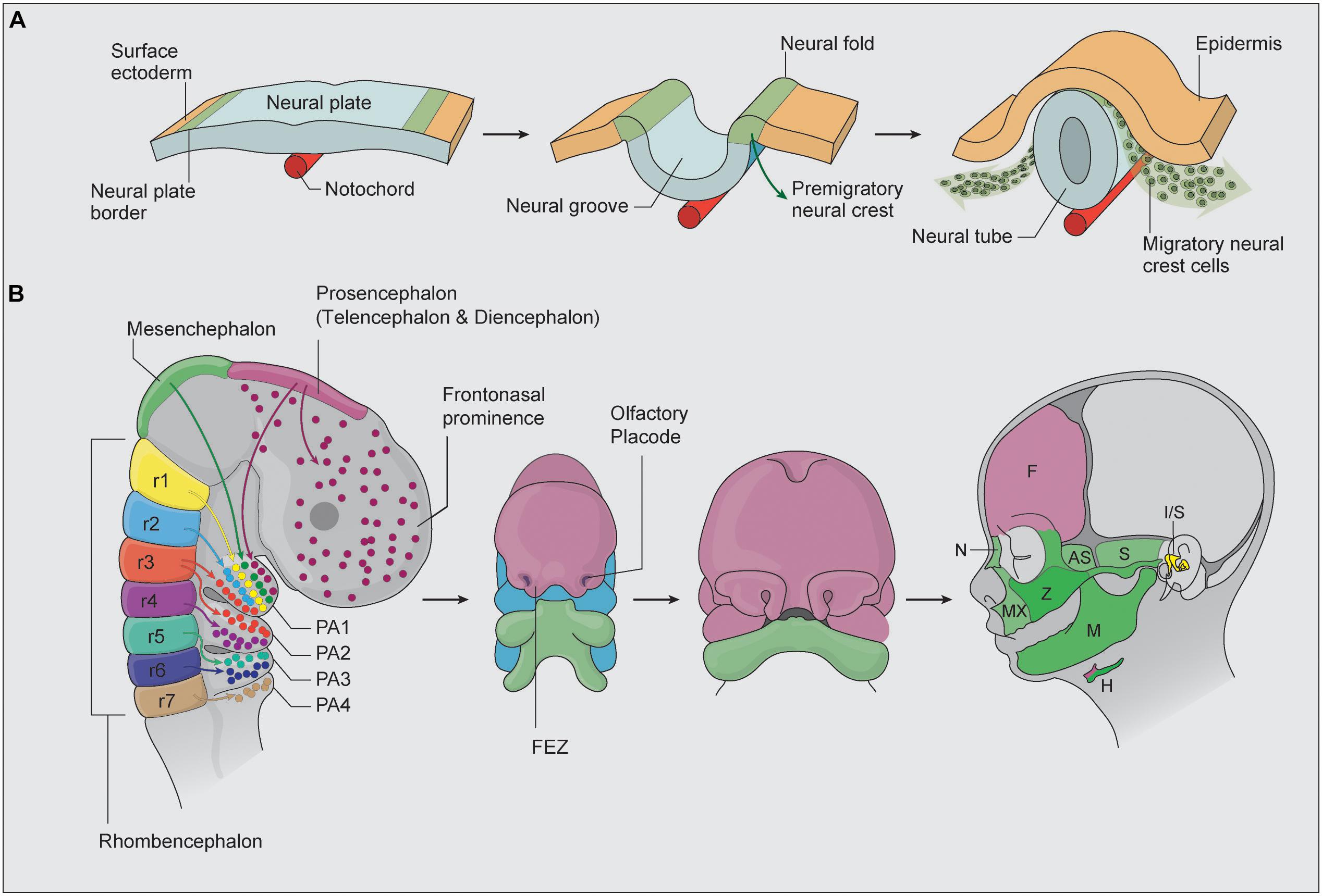
神経堤(NC: Neural Crest)は、一過性に現れる細胞群ですが、様々な細胞種に分化するため第四の胚葉とも言われています。脊椎動物では、神経堤細胞(NCC: Neural Crest Cell)神経管の背側隆起から剥離し、その後、神経形成中に発生中の胚内で広範囲に移動します1~3)。頭蓋神経堤細胞は咽頭弓に移動し、頭蓋顔面の大部分を構成し、最終的に歯、軟骨、頭蓋顔面骨、および結合組織を形成する間葉組織になります。 https://www.healthcare.nikon.com/ja/ss/cell-image-lab/glossary/neural-crest-cell.html
- 哺乳類の頭蓋骨発生の進化を解明(umut news:2014年4月7日) https://www.um.u-tokyo.ac.jp/information/news_20140407.html
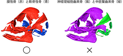
頭蓋冠は脳を覆 う ドーム状の骨組織で,主 として前頭骨, 頭頂骨, 後頭骨 に よ り構成 され る。頭蓋冠骨は膜性骨化によ り形成 され るが,そ の組織由来 は異なっており, 前頭骨は神経堤細胞由来, 頭頂骨と後頭骨は中胚葉由来であ る1)。
1) Jiang, X., Iseki, S., Maxson, R. E., Sucov, H. M. and Morriss-Kay, G. M.: Tissue origins and interaction in the mammalian skull vault. Dev. Biol. 241: 106-116, 2002. https://www.jstage.jst.go.jp/article/koubyou1952/71and72/4-1/71and72_4-1_19/_pdf/-char/ja
参考
- The squamous part of the temporal bone (or squamous temporalis/squamous temporal bone) is a very thin bone and forms the anterosuperior aspect of the temporal bone. https://radiopaedia.org/articles/squamous-part-of-temporal-bone
神経堤細胞から発生する構造
- Martik, M.L., Bronner, M.E. Riding the crest to get a head: neural crest evolution in vertebrates. Nat Rev Neurosci 22, 616–626 (2021). https://doi.org/10.1038/s41583-021-00503-2 無料要旨 In their seminal 1983 paper, Gans and Northcutt proposed that evolution of the vertebrate ‘new head’ was made possible by the advent of the neural crest and cranial placodes. The neural crest is a stem cell population that arises adjacent to the forming CNS and contributes to important cell types, including components of the peripheral nervous system and craniofacial skeleton and elements of the cardiovascular system. In the past few years, the new head hypothesis has been challenged by the discovery in invertebrate chordates of cells with some, but not all, characteristics of vertebrate neural crest cells.
- Diabetes, Oxidative Stress, and DNA Damage Modulate Cranial Neural Crest Cell Development and the Phenotype Variability of Craniofacial Disorders REVIEW article Front. Cell Dev. Biol., 20 May 2021 Sec. Molecular and Cellular Pathology Volume 9 – 2021 | https://doi.org/10.3389/fcell.2021.644410
- The Special Developmental Biology of Craniofacial Tissues Enables the Understanding of Oral and Maxillofacial Physiology and Diseases Int. J. Mol. Sci. 2021, 22(3), 1315; https://doi.org/10.3390/ijms22031315
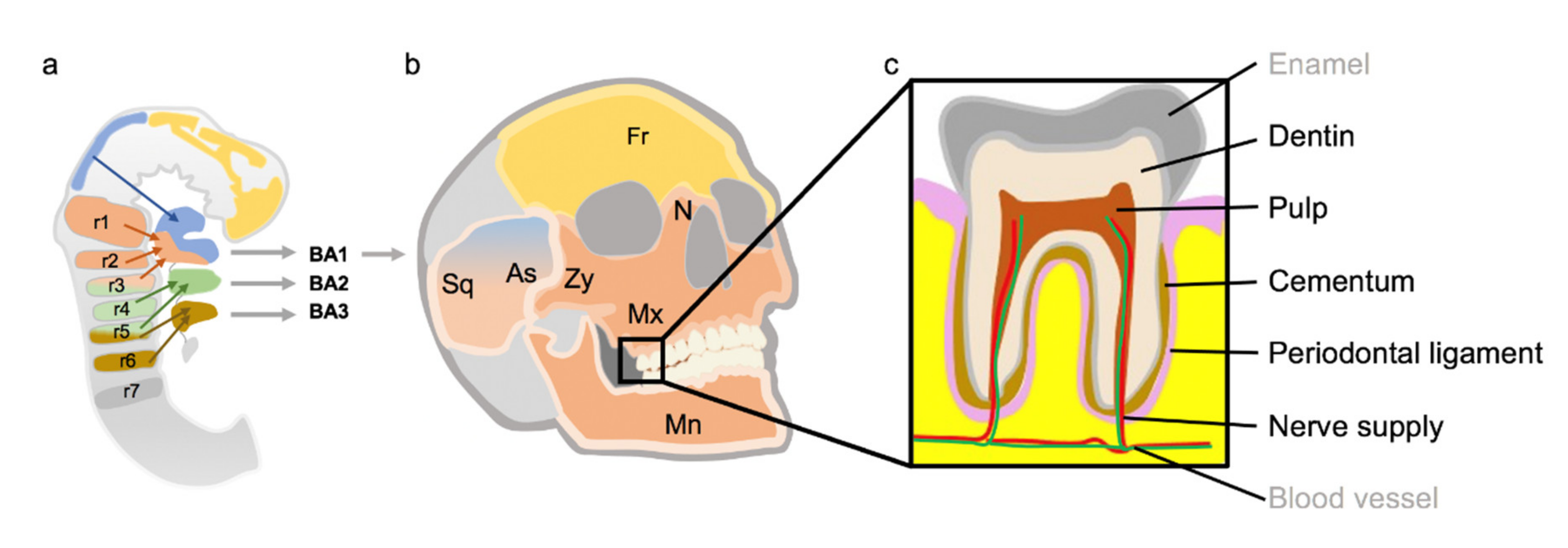
咽頭弓(鰓弓)から発生する顔面の構造
- Pharyngeal arches https://www.kenhub.com/en/library/anatomy/the-pharyngeal-arches
- First arch (mandibular) Skeletal structures, ligaments: Malleus 槌骨(つちこつ、ついこつ 中耳にあるハンマー(槌)の形をした小さい骨), short limb of incus キヌタ骨, maxilla 上顎骨, zygomatic bone 頬骨, hard palate, vomer bone 鋤骨, mandibule 下顎骨, temporal bone (squamous); anterior ligament of malleus, sphenomandibular ligament
- Second arch (hyoid) Skeletal structures, ligaments: Stapes 鐙骨(あぶみこつ 中耳にある骨で内耳へ音の振動を伝達する。) long limb of incus incus キヌタ骨(砧骨 音の振動を伝える中耳の三つの耳小骨の一つ), styloid process, lesser horn and upper part of body of hyoid bone 舌骨; stylohyoid ligament