発生の過程で胚の大きさが大きくなってくると、胚の隅々の細胞に酸素を供給する必要が出てきます。単細胞であれば酸素は拡散によって細胞内に取り込まれますが、胚の大きさが1㎜を超えるようになると奥の細胞は酸素が存在する外界と距離が遠すぎて物理的な拡散によって酸素を供給してもらうことは望めません。そのため、胚発生の早い段階で循環器系が機能する必要があります。種々の器官形成の中で、心臓の発生、機能の獲得が最も早いのも納得できます。
心臓になる細胞は、原始線条の時期に既に決まっています。原始線条のまわりにある細胞が内部に潜り込んで中胚葉となり、前方や側方に異動していって、さらに分化が進むわけですが、心臓になる細胞はまずPrimary Heart Fieldと呼ばれる側方の領域に左右一対としてあらわれます。それから領域が前方に伸びて左右が合わさり、一つのHeart Fieldtoなります。その時期のこの領域は、Heart Crescentと呼ばれます。
Heart field
Fig. 1. (A) Localization of heart precursors in the primary heart field during gastrulation and tubular heart formation. (B) Expression of BMP2, Nkx2.5, GATA5, and Tbx20 during an early stage (HH stage 5) of heart field formation in the chick embryo. https://www.sciencedirect.com/science/article/pii/S001216060300112X
Cardial Crescent
Cardial crescentはUの字のまがっている部分を指すので、構造としては1つのようです。しかし論文によっては、Uの字の両肩の曲がりに矢頭が2つあったりして、うっかりすると読み間違えそうになります。グーグルで”cardial crescents”で検索すると単数形に直されるので、やはり単数なのでしょう。

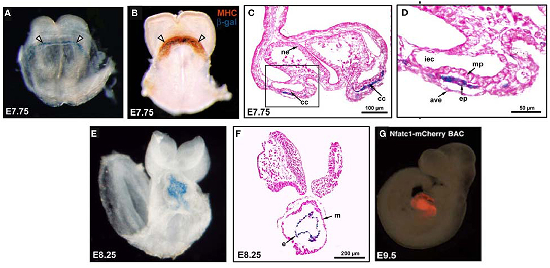
Figure 1. The endocardium and myocardium are intimately associated during early differentiation. (A) An NFATc1-nuc-LacZ embryo, E7.75, stained with X-Gal. Nuclear expression of β-gal (blue) within the endocardium of the cardiac crescent (white arrowheads). (B) An E7.75 NFATc1-nuc-LacZ embryo stained with myosin heavy chain (MHC; reddish-brown) to identify the myocardium and co-stained with X-Gal to mark β-gal+ (blue) endocardium within the cardiac crescent (white arrowheads).
Cardial CrescentとEndocardial tubesの時期
発生学の教科書やネットの説明、レビュー論文の説明をいろいろみていると、左右一対のHeart fieldが形成されその前方部分が伸びて繋がってhear Crescentと呼ばれる領域になり心臓が形成されると説明しているものと、左右一対のendocardial tubesが生じて、真ん中にきて融合して一本のチューブになると説明しているものと2種類を良くみかけます。すると、Uの字を逆向きにしたようなCrescentからなぜ2本の管になるのか、が理解できませんでした。そもそもどっちのステージが先なのかすら曖昧に感じていました。ChatGPTに訊くと、Crescentの形成が先のようです。明解な説明を探していますが、見当たらないのが不思議です。
心臓の発生
https://www.researchgate.net/figure/Mouse-embryos-showing-major-stages-of-heart-development-A-C-Ventral-view-of-mouse_fig2_254851589
