哺乳類の受精卵が卵割を繰り返したあと、最初に「分化」でできるのは、内部細胞塊と栄養層です。この2つの細胞の種類はどのようにして分化するのでしょうか。
最初のこの分化に関与するのはHIPPOシグナリングと呼ばれるリン酸化の反応系です。HIPPOという名前はショウジョウバエの研究で名付けられたもので、哺乳類ではHIPPOはMST1,MST2と呼ばれます。HIPPO(MST1/2)は、キナーゼカスケードシグナリングの上流に位置するキナーゼです。HIPPOがどうのようにして活性化されるのかは、実は多様性があり、細胞同士の接触、細胞に対するストレス、細胞外からやってくる成長因子に対する受容体を介するものがあります。
Hippo Signal Pathway Creative BioMart チャンネル登録者数 3220人 チャンネル登録
HIPPO(カバの意味)の役割は増殖を止めることだそうです。
Hippo Signaling Regulates Microprocessor and Links Cell-Density-Dependent miRNA Biogenesis to Cancer Cell Press チャンネル登録者数 2.55万人
もともとの発見は、ショウジョウバエで組織の細胞増殖に異常を示す突然変異体として同定されました。
- Sci Rep. 2020; 10: 3173. Published online 2020 Feb 21. doi: 10.1038/s41598-020-60120-4 PMCID: PMC7035326 PMID: 32081887 Systematic analysis of the Hippo pathway organization and oncogenic alteration in evolution
- The discovery and expansion of Hippo signaling pathway in Drosophila model July 2017Hereditas (Beijing) 39(7):537-545 39(7):537-545 DOI:10.16288/j.yczz.17-051 https://www.researchgate.net/publication/320893314_The_discovery_and_expansion_of_Hippo_signaling_pathway_in_Drosophila_model 本文は中国語? The discovery of Hippo signaling pathway is another breakthrough of fly genetics. Similar to the other signaling pathways, Hippo pathway also functions crucially in tremendous physiological and pathological conditions, like organ size control and cancer. There are three main stages of Hippo pathway study: Firstly, identifications of core components by fly genetic screens; secondly, regulations by versatile upstream cues, like cytoskeleton, mechanical tension, and nutrition
HIPPOシグナリングを構成するシグナル分子
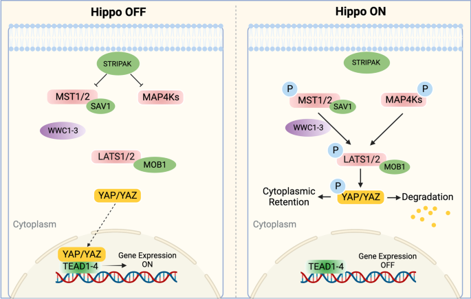
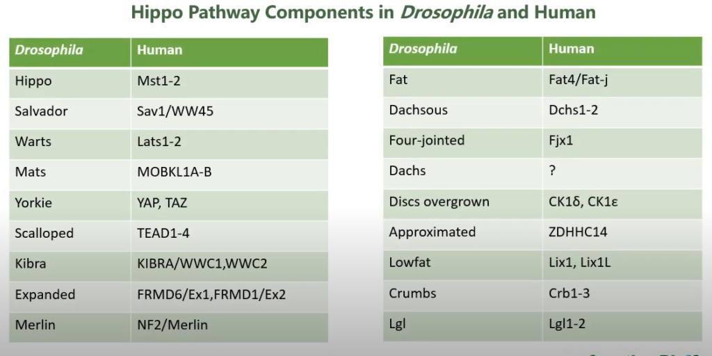
In mammals, the Hippo pathway is composed of several key components, including mammalian STE20-like kinase 1/2 (MST1/2), protein Salvador homologue 1 (SAV1), MOBKL1A/B (MOB1A/B), large tumour suppressor kinase 1/2 (LATS1/2), Yes-associated protein 1 (YAP), WW-domain-containing transcription regulator 1 (TAZ), and the transcriptional enhanced associated domain (TEAD) family1 (Fig. 2). YAP/TAZ are transcriptional coactivators that bind to TEAD1–4 to regulate the expression of a wide array of genes that mediate cell proliferation, apoptosis, and stem cell self-renewal.2 Moreover, a variety of upstream signals, such as cell polarity, mechanical cues, cell density, soluble factors and stress signals, modulate the Hippo pathway.3,4,5
08 November 2022 The Hippo signalling pathway and its implications in human health and diseases https://www.nature.com/articles/s41392-022-01191-9 オープンアクセス論文
HIPPOシグナルのON/OFFとシグナリングの効果
HIPPOキナーゼが活性化された状態(リン酸化された状態)の場合は、HIPPOキナーゼはLATSキナーゼをリン酸化し、今度はLATSキナーゼがYAPをリン酸化することで、YAPが核内に移行できなくなり、細胞質中に保持されて分解されてしまいます。それに対して、HIPPOキナーゼが活性かされていないときは、MSTにキナーゼ活性がないため、YAPはリン酸化されておらず、核内に移行できて、転写因子TEAD4と結合して、転写因子として標的遺伝子の発現を促します。
 https://www.nature.com/articles/s41392-022-01191-9
ショウジョウバエにおけるHIPPOシグナリング
The central core of this pathway includes a pair of kinases, Hippo and Warts (Wts), which act in sequence to phosphorylate the transcriptional co-activator Yorkie (Yki) (Huang et al., 2005). Phosphorylation of Yki by Wts promotes cytoplasmic localization of Yki, thus reducing Yki-dependent transcription and growth (Dong et al., 2007; Oh and Irvine, 2008). https://www.ncbi.nlm.nih.gov/pmc/articles/PMC6215397/
 出典:https://www.youtube.com/watch?v=4qfj53CFIDU 6分37秒頃
哺乳類のHIPPOシグナリングの複雑さ
The mammalian Hippo pathway is more complicated than the Drosophila Hippo pathway. One of the reasons for this complexity is that mammals have more than one paralogue for each Drosophila component. These paralogues sometimes play redundant roles but in most cases exhibit distinct properties. Second and more importantly, the components of the mammalian Hippo pathway undergo many molecular interactions, so they exert additional functions and are subject to additional regulation. For instance, the substrates of MST kinases include not only LATS kinases and MOB1, but also c-Jun N-terminal kinase (JNK), histone H2B and FoxO, as discussed below (85,86). All of them are implicated in apoptosis. LATS1 interacts with LIM domain kinase 1 to inhibit its kinase activity and thereby affects cytokinesis (87). It also binds mitochondrial serine protease Omi/HtrA2 to promote the protease activity (88,89). Omi/HtrA2 controls cell proliferation through LATS1. If we define the final outputs of the Hippo pathway as the regulation of cell proliferation and cell death, it can be argued that these molecular interactions also mediate Hippo signalling. No matter how we demarcate the Hippo pathway, we need to consider that activation of the MST–LATS–YAP/TAZ axis is associated with parallel activation of other pathways, which co-operate with the canonical Hippo pathway. https://academic.oup.com/jb/article/149/4/361/968447
Trophoblastへの分化
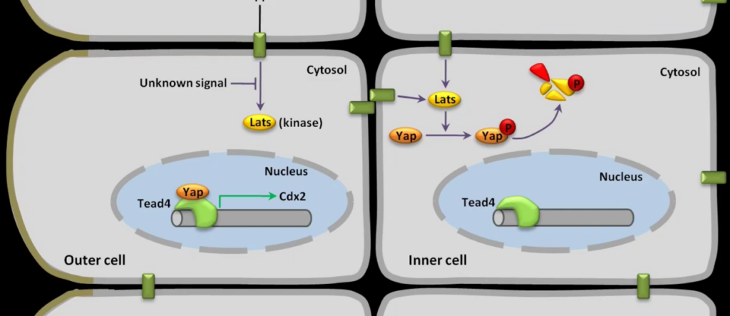
下の動画ではHIPPOシグナリングがなぜTrophoblastとInner Cell Massの2種類に分化誘導するかそのメカニズムを説明しています。HIPPOを抑制する因子が外側の細胞では何なのかについては不明としています。
 Possible pathway initiating the distinction between inner cell mass and trophoblast Bio peak チャンネル登録者数 2480人 https://www.youtube.com/watch?v=bwcICTcF2wE